Video: Every Case Tells a Story| Webinar: ACR/CHEST ILD Guidelines in Practice

An official publication of the ACR and the ARP serving rheumatologists and rheumatology professionals

  • Conditions
    • Axial Spondyloarthritis
    • Gout and Crystalline Arthritis
    • Myositis
    • Osteoarthritis and Bone Disorders
    • Pain Syndromes
    • Pediatric Conditions
    • Psoriatic Arthritis
    • Rheumatoid Arthritis
    • Sjögren’s Disease
    • Systemic Lupus Erythematosus
    • Systemic Sclerosis
    • Vasculitis
    • Other Rheumatic Conditions
  • FocusRheum
    • ANCA-Associated Vasculitis
    • Axial Spondyloarthritis
    • Gout
    • Psoriatic Arthritis
    • Rheumatoid Arthritis
    • Systemic Lupus Erythematosus
  • Guidance
    • Clinical Criteria/Guidelines
    • Ethics
    • Legal Updates
    • Legislation & Advocacy
    • Meeting Reports
      • ACR Convergence
      • Other ACR meetings
      • EULAR/Other
    • Research Rheum
  • Drug Updates
    • Analgesics
    • Biologics/DMARDs
  • Practice Support
    • Billing/Coding
    • EMRs
    • Facility
    • Insurance
    • QA/QI
    • Technology
    • Workforce
  • Opinion
    • Patient Perspective
    • Profiles
    • Rheuminations
      • Video
    • Speak Out Rheum
  • Career
    • ACR ExamRheum
    • Awards
    • Career Development
  • ACR
    • ACR Home
    • ACR Convergence
    • ACR Guidelines
    • Journals
      • ACR Open Rheumatology
      • Arthritis & Rheumatology
      • Arthritis Care & Research
    • From the College
    • Events/CME
    • President’s Perspective
  • Search

Diagnosing Anti-MOG Autoimmune Encephalomyelitis

Thomas R. Collins  |  April 6, 2020

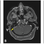
ATLANTA—A 7-year-old girl presented to her pediatrician with a fever, nausea, vomiting and a headache, and was diagnosed with a sinus infection. As her symptoms grew worse and more complex, the case led neurologists and rheumatologists on a long hunt for a diagnosis—ultimately, anti-myelin oligodendrocyte glycoprotein (anti-MOG) autoimmune encephalomyelitis—that highlighted how patience, a thorough workup and multi-department teamwork can be crucial for proper diagnosis and management of a pediatric patient with inflammatory brain disease.

A discussion of the case and its lessons was led at the 2019 ACR/ARP Annual Meeting by Elizabeth Wells, MD, director of inpatient neurology at Children’s National Hospital, Washington, D.C., and Heather Van Mater, MD, MSc, a pediatric rheumatologist at Duke Children’s Hospital, Durham, N.C.

ad goes here:advert-1
ADVERTISEMENT
SCROLL TO CONTINUE

Initial Diagnosis
After the sinus infection diagnosis, the girl’s headache got worse and she had difficulty using her hand, prompting an emergency department visit. She was found to have sinusitis on a computerized tomography scan, with a white blood count (WBC) of 12.5 and a C-reactive protein (CRP) level of 1.6. With a severe headache, a moody disposition and sleepiness at Day 12, she was referred to neurology.

“The biggest things for me as a neurologist at this point would be that focal complaint—that her right hand was not working at some point—as well as the fact that the headache is severe, persistent and not getting better—and that it was her first headache,” with no history of headache or migraine, Dr. Wells said.

ad goes here:advert-2
ADVERTISEMENT
SCROLL TO CONTINUE

A magnetic resonance imaging (MRI) scan showed sphenoid sinusitis with T2 changes in the frontal lobe. Because of the migraine, she was given prednisone for five days on day 17. Her symptoms resolved, but after the steroids were stopped she again developed a headache and a low-grade fever, with excessive sleepiness and irritability.

She was admitted to the hospital on the 25th day, still with a presumed migraine. She had a CRP of 11.7, WBC of 34.9 and hemoglobin of 12.9. She was treated for migraine, released with an oral steroid taper and improved at home.

Signs of a Deeper Problem Missed
Dr. Wells said signs of a deeper problem were missed early on.

“There may have been some anchoring bias here, where she got diagnosed with migraine and then was getting better, but I still think back to those focal symptoms she had initially,” as well as her abnormal lab findings, Dr. Wells said.

A few days later, things got much worse. The girl had hallucinations and a seizure that became status epilepticus, with a WBC of 34.8, an erythrocyte sedimentation rate (ESR) of 36 and a CRP of 11.2. A lumbar puncture found a WBC of 211 with a segmented neutrophil reading of 81% and red blood cell count of zero.

Page: 1 2 3 4 | Single Page
Share: 

Filed under:ConditionsPediatric Conditions Tagged with:anti-myelin oligodendrocyte glycoprotein (anti-MOG) autoimmune encephalomyelitisbrainChildrenencephalomyelitisPediatric

Related Articles

    APS: What Rheumatologists Should Know about Hughes Syndrome

    February 17, 2016

    The problem that dogs the work of all of those treating patients with antiphospholipid syndrome (APS) is the apparent lack of knowledge of the syndrome, both by the general public, as well as by swaths of the medical fraternity. Perhaps it was ever thus—a syndrome less than 40 years old could be described as new,…

    Case Report: A Behçet’s Patient Develops Cerebral Venous Sinus Thrombi

    December 17, 2018

    A 39-year-old woman presented at the emergency department with three weeks of progressive, constant and pulsatile right-sided headache. She said her headache was worse in the morning and when she would bend forward. She reported associated nausea and vomiting. On initial assessment, she did not have any focal neurological deficits. Her medical history was significant for…

    Can Systemic Inflammation Influence Mood?

    August 17, 2015

    The Friday night press release: When a politician or any public figure needs to disclose unfavorable news, chances are they will release it sometime on a late Friday afternoon or evening, hoping that nobody is paying attention. In fact, this behavior was coined “the take out the trash day” on the television political drama, The…

    Cryopyrin-Associated Periodic Syndromes: Difficult to Recognize, Diagnose, Treat

    October 1, 2014

    Two case studies demonstrate the difficulty, delay in recognizing this rare autoinflammatory disease

  • About Us
  • Meet the Editors
  • Issue Archives
  • Contribute
  • Advertise
  • Contact Us
  • Copyright © 2025 by John Wiley & Sons, Inc. All rights reserved, including rights for text and data mining and training of artificial technologies or similar technologies. ISSN 1931-3268 (print). ISSN 1931-3209 (online).
  • DEI Statement
  • Privacy Policy
  • Terms of Use
  • Cookie Preferences