Editor’s note: In this recurring feature, we first present a series of images (this page) for your review, and then a brief discussion of the findings and diagnosis. Before you turn to the discussion, examine these images carefully and draw your own conclusions.

Dr. Leatherwood

Dr. Todd
History
A 33-year-old woman with a 16-year history of systemic lupus erythematosus (SLE) reports chronic bilateral foot pain and deformity. Serologic studies include an anti-nuclear antibody (ANA) at 1:5,120 titer (diffuse pattern), with very elevated anti-double-stranded DNA (dsDNA) and anti-ribonucleoprotein (RNP) antibody titers. Serum C3 and C4 complement levels have been chronically low. SLE features have consisted primarily of mucocutaneous, articular, and serositis disease. The following radiographic images are obtained.
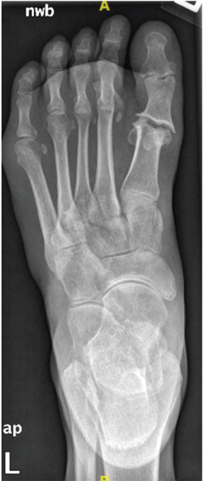
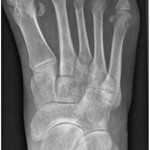
Figure 1: Anteroposterior (AP) radiograph of the left foot.
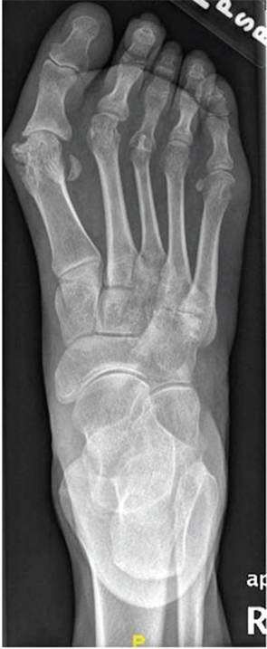
Figure 2: AP radiograph of the right foot.