Gout arthritis can occur in any joint of the body. As in this case, patients with chronic renal insufficiency frequently have hyperuricemia and may develop gout, often in uncommon locations. Although the imaging findings are fairly characteristic, with well-defined marginal erosions with overhanging edges and relative preservation of the joint space until the late stages of disease, biopsy and/or aspiration may be indicated to confirm the diagnosis.
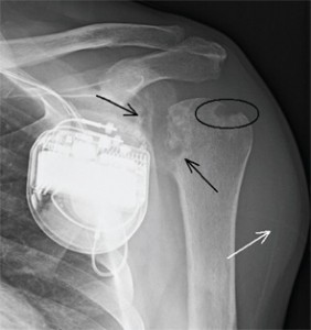
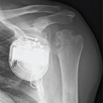
Figure 1: AP radiograph of the left shoulder.
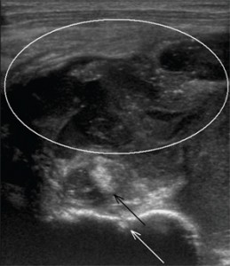
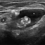
Figure 2: Transverse sonographic image of the left shoulder.
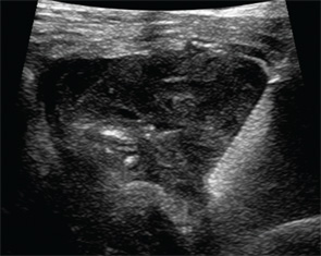
Figure 3: Transverse sonographic image of the left shoulder during aspiration and biopsy.
Jennifer L. Demertzis, MD, is a musculoskeletal radiologist at the Mallinckrodt Institute of Radiology at Washington University School of Medicine in St. Louis, Mo. She is excited to collaborate on this new feature in the journal and looks forward to seeing future cases contributed by readers.
Send Us Your Images
Contact us at:
Keri Losavio
editor
E-mail: [email protected]