For the discussion, click here.
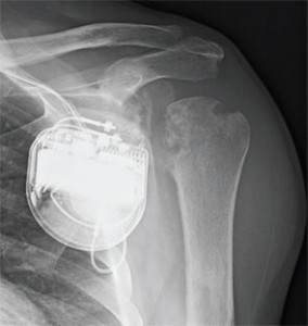
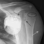
Figure 1: Anteroposterior (AP) radiograph of the left shoulder.
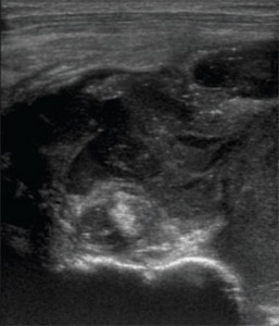
Figure 2: Transverse sonographic image of the left shoulder at the level of the proximal humerus and extra-articular long-head biceps tendon.
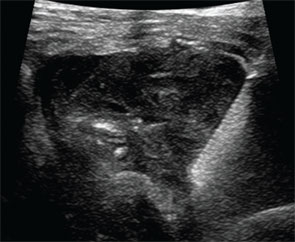
Figure 3: Transverse sonographic image of the left shoulder during image-guided aspiration and biopsy from an anterolateral approach (needle on the right side of the image).