The Rheumatologist
COVID-19 News
  • Connect with us:
  • Facebook
  • Twitter
  • LinkedIn
  • YouTube
  • Feed
  • Home
  • Conditions
    • Rheumatoid Arthritis
    • SLE (Lupus)
    • Crystal Arthritis
      • Gout Resource Center
    • Spondyloarthritis
    • Osteoarthritis
    • Soft Tissue Pain
    • Scleroderma
    • Vasculitis
    • Systemic Inflammatory Syndromes
    • Guidelines
  • Resource Centers
    • Ankylosing Spondylitis Resource Center
    • Gout Resource Center
    • Rheumatoid Arthritis Resource Center
    • Systemic Lupus Erythematosus Resource Center
  • Drug Updates
    • Biologics & Biosimilars
    • DMARDs & Immunosuppressives
    • Topical Drugs
    • Analgesics
    • Safety
    • Pharma Co. News
  • Professional Topics
    • Ethics
    • Legal
    • Legislation & Advocacy
    • Career Development
      • Certification
      • Education & Training
    • Awards
    • Profiles
    • President’s Perspective
    • Rheuminations
  • Practice Management
    • Billing/Coding
    • Quality Assurance/Improvement
    • Workforce
    • Facility
    • Patient Perspective
    • Electronic Health Records
    • Apps
    • Information Technology
    • From the College
    • Multimedia
      • Audio
      • Video
  • Resources
    • Issue Archives
    • ACR Convergence
      • Systemic Lupus Erythematosus Resource Center
      • Rheumatoid Arthritis Resource Center
      • Gout Resource Center
      • Abstracts
      • Meeting Reports
      • ACR Convergence Home
    • American College of Rheumatology
    • ACR ExamRheum
    • Research Reviews
    • ACR Journals
      • Arthritis & Rheumatology
      • Arthritis Care & Research
      • ACR Open Rheumatology
    • Rheumatology Image Library
    • Treatment Guidelines
    • Rheumatology Research Foundation
    • Events
  • About Us
    • Mission/Vision
    • Meet the Authors
    • Meet the Editors
    • Contribute to The Rheumatologist
    • Subscription
    • Contact
  • Advertise
  • Search
You are here: Home / Articles / Diagnostic Imaging in Patient with Elbow Pain: Findings

Diagnostic Imaging in Patient with Elbow Pain: Findings

May 14, 2015 • By Veronika Sharp, MD, Midori Jane Nishio, MD, & Lily Kao, MD

  • Tweet
  • Email
Print-Friendly Version / Save PDF
Figure 1

Figure 1

View the question.

You Might Also Like
  • Diagnostic Imaging in Patient with Elbow Pain: History
  • Diagnostic Imaging in Patient with Atraumatic Left Shoulder Pain: Findings
  • Diagnostic Imaging in Patient with Atraumatic Left Shoulder Pain: History
Explore This Issue
May 2015

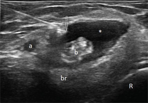
Transverse (see Figure 1) and longitudinal (see Figure 2) ultrasound images show an anechoic collection (asterisks),

ad goes here:advert-1
ADVERTISEMENT
SCROLL TO CONTINUE

which surrounds the distal biceps tendon (b). The collection is compressible (not shown here). The biceps tendon is heterogeneous in echo texture, with fiber dropouts and an irregular border (in the transverse view). The elbow joint does not show abnormalities (images not shown); hence, the collection was not felt to communicate with the joint. Aspiration of the collection was performed under ultrasound guidance (needle tip: arrow).

The ultrasound findings are consistent with bicipitoradial bursitis and distal bicep tendinosis. The distal biceps tendon is not covered by a synovial sheath, but rather by paratenon. Just proximal to its insertion to the radial tuberosity, the biceps tendon is in contact with the bicipitoradial (cubital) bursa, which does not communicate with the elbow joint but when distended can completely surround the biceps tendon and mimic tenosynovitis. Bicipitoradial bursitis should be distinguished from ganglion cysts, which may arise from the anterior elbow joint, connected via a pedicle, and expand distally.

ad goes here:advert-2
ADVERTISEMENT
SCROLL TO CONTINUE

Bicipitoradial bursitis can result from repetitive mechanical trauma, overuse, distal biceps tendon tear, tuberculosis,
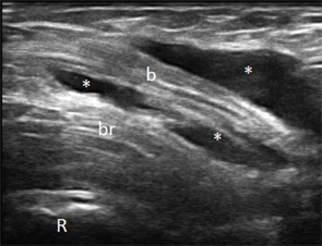
Figure 2

Figure 2

inflammatory arthritis, synovial chrondromatosis or amyloidosis. In our case the fluid was noninflammatory and crystal negative. Gram stain and culture, AFB smear and culture, cytology and Congo red stain were negative. The most likely reason for the bursitis remained overuse. Her symptoms improved with steroid injection and rest. Rarely, severe cases of bicipitoradial bursitis may require surgical resection of the bursa.

Ultrasound is useful in identifying bursitis, provides guidance for aspiration and injection, and allows for assessment of bicep tendon tear and macroscopic radial nerve damage resulting from compression.

ad goes here:advert-3
ADVERTISEMENT
SCROLL TO CONTINUE

Veronika Sharp, MD, is a faculty rheumatologist at the Santa Clara Valley Medical Center (SCVMC) in San Jose and is an affiliated clinical assistant professor at Stanford University, Palo Alto, Calif. She is director of musculoskeletal (MSK) ultrasonography in rheumatology at SCVMC.

Midori Jane Nishio, MD, is a rheumatologist in Walnut Creek, Calif., and is a volunteer faculty at SCVMC in MSK ultrasonography.

Lily Kao, MD, is a rheumatologist in San Jose, Calif., and is a volunteer faculty at SCVMC in MSK ultrasonography.


Send Us Your Images

Contact us at:

Simon Helfgott, MD
physician editor
E-mail: shelfgott@partners.org

Keri Losavio
editor
E-mail: klosavio@wiley.com

Filed Under: Conditions Tagged With: bursitis, imaging, Joint Pain, rheumatologistIssue: May 2015

You Might Also Like:
  • Diagnostic Imaging in Patient with Elbow Pain: History
  • Diagnostic Imaging in Patient with Atraumatic Left Shoulder Pain: Findings
  • Diagnostic Imaging in Patient with Atraumatic Left Shoulder Pain: History
  • Diagnostic Imaging in Lupus Patient with Foot Pain: Findings

Meeting Abstracts

Browse and search abstracts from the ACR Convergence and ACR/ARP Annual Meetings going back to 2012.

Visit the Abstracts site »

American College of Rheumatology

Visit the official website for the American College of Rheumatology.

Visit the ACR »

ACR Convergence

Don’t miss rheumatology’s premier scientific meeting for anyone involved in research or the delivery of rheumatologic care or services.

Visit the ACR Convergence site »

The Rheumatologist newsmagazine reports on issues and trends in the management and treatment of rheumatic diseases. The Rheumatologist reaches 11,500 rheumatologists, internists, orthopedic surgeons, nurse practitioners, physician assistants, nurses, and other healthcare professionals who practice, research, or teach in the field of rheumatology.

About Us / Contact Us / Advertise / Privacy Policy / Terms of Use

  • Connect with us:
  • Facebook
  • Twitter
  • LinkedIn
  • YouTube
  • Feed

Copyright © 2006–2021 American College of Rheumatology. All rights reserved.

ISSN 1931-3268 (print)
ISSN 1931-3209 (online)

loading Cancel
Post was not sent - check your email addresses!
Email check failed, please try again
Sorry, your blog cannot share posts by email.
This site uses cookies: Find out more.