The Rheumatologist
COVID-19 News
  • Connect with us:
  • Facebook
  • Twitter
  • LinkedIn
  • YouTube
  • Feed
  • Home
  • Conditions
    • Rheumatoid Arthritis
    • SLE (Lupus)
    • Crystal Arthritis
      • Gout Resource Center
    • Spondyloarthritis
    • Osteoarthritis
    • Soft Tissue Pain
    • Scleroderma
    • Vasculitis
    • Systemic Inflammatory Syndromes
    • Guidelines
  • Resource Centers
    • Ankylosing Spondylitis Resource Center
    • Gout Resource Center
    • Rheumatoid Arthritis Resource Center
    • Systemic Lupus Erythematosus Resource Center
  • Drug Updates
    • Biologics & Biosimilars
    • DMARDs & Immunosuppressives
    • Topical Drugs
    • Analgesics
    • Safety
    • Pharma Co. News
  • Professional Topics
    • Ethics
    • Legal
    • Legislation & Advocacy
    • Career Development
      • Certification
      • Education & Training
    • Awards
    • Profiles
    • President’s Perspective
    • Rheuminations
  • Practice Management
    • Billing/Coding
    • Quality Assurance/Improvement
    • Workforce
    • Facility
    • Patient Perspective
    • Electronic Health Records
    • Apps
    • Information Technology
    • From the College
    • Multimedia
      • Audio
      • Video
  • Resources
    • Issue Archives
    • ACR Convergence
      • Systemic Lupus Erythematosus Resource Center
      • Rheumatoid Arthritis Resource Center
      • Gout Resource Center
      • Abstracts
      • Meeting Reports
      • ACR Convergence Home
    • American College of Rheumatology
    • ACR ExamRheum
    • Research Reviews
    • ACR Journals
      • Arthritis & Rheumatology
      • Arthritis Care & Research
      • ACR Open Rheumatology
    • Rheumatology Image Library
    • Treatment Guidelines
    • Rheumatology Research Foundation
    • Events
  • About Us
    • Mission/Vision
    • Meet the Authors
    • Meet the Editors
    • Contribute to The Rheumatologist
    • Subscription
    • Contact
  • Advertise
  • Search
You are here: Home / Articles / Diagnostic Imaging in Patient with Chronic Left Ankle Pain: Findings

Diagnostic Imaging in Patient with Chronic Left Ankle Pain: Findings

April 15, 2016 • By Cianna Leatherwood, MD, & Derrick J. Todd, MD, PhD

  • Tweet
  • Email
Print-Friendly Version / Save PDF

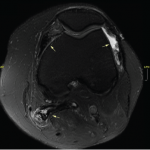
Radiographic imaging showed circumferential soft tissue swelling of the ankle with a soft-tissue density seen in the tibiotalar and posterior subtalar joints, as well as a large, lobulated effusion. MRI of the left ankle shows cystic changes within the talus and first cuneiform bones, as well as a lobulated abnormal soft tissue density with low signal intensity within the tibiotalar and subtalar joints. The MRI suggested a diagnosis of pigmented villonodular synovitis (PVNS) with associated subchondral erosions, a diagnosis that was confirmed by ultrasound-guided synovial tissue biopsy.

You Might Also Like
  • Diagnostic Imaging in Patient with Chronic Left Ankle Pain: History
  • Diagnostic Imaging in Patient with Left Hip Pain: Findings
  • The Diagnostic View: Our Findings
Explore This Issue
April 2016
Also By This Author
  • Diagnostic Imaging in Patient with Chronic Left Ankle Pain: History

PVNS is an uncommon disorder of unknown etiology, affecting synovial and tenosynovial tissues. It is more common in women than in men. There are two major types of PVNS: diffuse and localized. Diffuse PVNS presents as indolent and progressive enlargement of the entirety of a single joint. The knee is the most commonly affected joint, followed by the ankle and the hip. When PVNS affects the tendon sheath of a digit, it is often referred to as a tenosynovial giant cell tumor. Rarely, PVNS can affect the spine. Localized PVNS occurs less frequently than the diffuse PVNS, and affects only a portion of the synovium in one joint. The affected portion can project into the joint space, which can cause a patient to experience a foreign body sensation, such as locking, clicking or catching. Pain is less common in localized PVNS. Aspiration of any effusion associated with PVNS classically produces dark, hemosiderin laden fluid. The appearance of such fluid in the absence of antecedent trauma should prompt consideration of the presence of PVNS.

ad goes here:advert-1
ADVERTISEMENT
SCROLL TO CONTINUE

Often, radiographs will not identify PVNS. Non-contrast MRI will show a dark, soft tissue mass with low-to-intermediate signal intensity on T1-weighted imaging, and low signal intensity on T2-weighted imaging, as a result of hemosiderin deposition in synovial tissue. Additional findings of scalloped erosions with sclerotic changes at sites of synovial insertion can be seen in cases of prolonged PVNS.

Treatment of localized and diffuse PVNS is surgical resection of the lesion and affected synovium. Without complete synovial resection, recurrences occur frequently. Adjunctive intra-articular radiation or external beam radiation therapy may reduce the risk of PVNS recurrence, especially in cases of diffuse disease refractory to initial surgical resection. In cases of advanced diffuse PVNS, when there is invasive destruction of bone, the gold standard of treatment is complete synovectomy with total joint arthroplasty. Vertebral involvement of PVNS can be difficult to treat with surgery. There are a few reports of successful treatment of PVNS with the tyrosine kinase inhibitor imatinib mesylate or anti-TNF agents.

ad goes here:advert-2
ADVERTISEMENT
SCROLL TO CONTINUE

Pages: 1 2 | Single Page

Filed Under: Conditions, Soft Tissue Pain Tagged With: ankle, diagnostic, foot, imaging, MRI, Pain, radiography, rheumatology, swellingIssue: April 2016

You Might Also Like:
  • Diagnostic Imaging in Patient with Chronic Left Ankle Pain: History
  • Diagnostic Imaging in Patient with Left Hip Pain: Findings
  • The Diagnostic View: Our Findings
  • Diagnostic Imaging in Lupus Patient with Foot Pain: Findings

Simple Tasks

Learn more about the ACR’s public awareness campaign and how you can get involved. Help increase visibility of rheumatic diseases and decrease the number of people left untreated.

Visit the Simple Tasks site »

ACR Convergence

Don’t miss rheumatology’s premier scientific meeting for anyone involved in research or the delivery of rheumatologic care or services.

Visit the ACR Convergence site »

American College of Rheumatology

Visit the official website for the American College of Rheumatology.

Visit the ACR »

The Rheumatologist newsmagazine reports on issues and trends in the management and treatment of rheumatic diseases. The Rheumatologist reaches 11,500 rheumatologists, internists, orthopedic surgeons, nurse practitioners, physician assistants, nurses, and other healthcare professionals who practice, research, or teach in the field of rheumatology.

About Us / Contact Us / Advertise / Privacy Policy / Terms of Use

  • Connect with us:
  • Facebook
  • Twitter
  • LinkedIn
  • YouTube
  • Feed

Copyright © 2006–2021 American College of Rheumatology. All rights reserved.

ISSN 1931-3268 (print)
ISSN 1931-3209 (online)

loading Cancel
Post was not sent - check your email addresses!
Email check failed, please try again
Sorry, your blog cannot share posts by email.
This site uses cookies: Find out more.