Editor’s note: In this recurring feature, we first present a series of images (this page) for your review, and then a brief discussion of the findings and diagnosis. Before you turn to the discussion, examine these images carefully and draw your own conclusions.
History
A 49-year-old woman presents with one year of chronic left ankle pain four years after she sprained the same ankle. She participated in physical therapy after the initial sprain and experienced three years of relief from ankle pain and swelling. However, her symptoms returned one year prior to her current presentation. The pain and swelling returned gradually and increased over time. A radiograph at the time was interpreted as showing swelling of the ankle but was otherwise normal. She received an injection of corticosteroids six months prior to the current presentation, without improvement in pain or swelling. Radiography and non-contrast enhanced MRI of the left ankle revealed the following images.
For the discussion, click here.
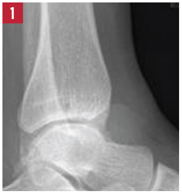
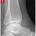
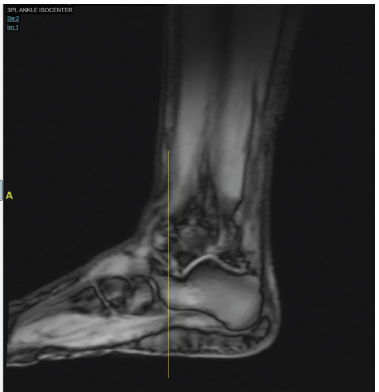
X-ray of the left ankle.
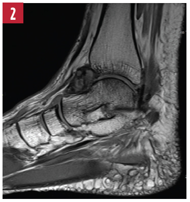
T1-weighted image of the left ankle.
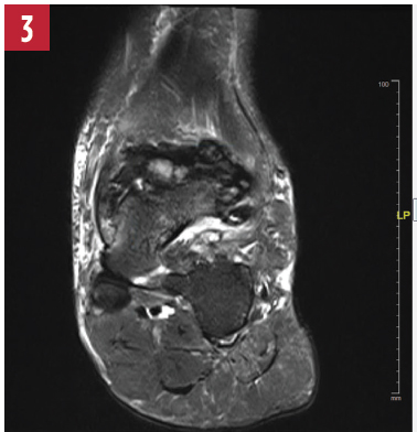
T2-weighted image of the left ankle.
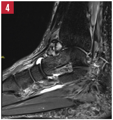
STIR image of the left ankle.