Video: Every Case Tells a Story| Webinar: ACR/CHEST ILD Guidelines in Practice

An official publication of the ACR and the ARP serving rheumatologists and rheumatology professionals

  • Conditions
    • Axial Spondyloarthritis
    • Gout and Crystalline Arthritis
    • Myositis
    • Osteoarthritis and Bone Disorders
    • Pain Syndromes
    • Pediatric Conditions
    • Psoriatic Arthritis
    • Rheumatoid Arthritis
    • Sjögren’s Disease
    • Systemic Lupus Erythematosus
    • Systemic Sclerosis
    • Vasculitis
    • Other Rheumatic Conditions
  • FocusRheum
    • ANCA-Associated Vasculitis
    • Axial Spondyloarthritis
    • Gout
    • Psoriatic Arthritis
    • Rheumatoid Arthritis
    • Systemic Lupus Erythematosus
  • Guidance
    • Clinical Criteria/Guidelines
    • Ethics
    • Legal Updates
    • Legislation & Advocacy
    • Meeting Reports
      • ACR Convergence
      • Other ACR meetings
      • EULAR/Other
    • Research Rheum
  • Drug Updates
    • Analgesics
    • Biologics/DMARDs
  • Practice Support
    • Billing/Coding
    • EMRs
    • Facility
    • Insurance
    • QA/QI
    • Technology
    • Workforce
  • Opinion
    • Patient Perspective
    • Profiles
    • Rheuminations
      • Video
    • Speak Out Rheum
  • Career
    • ACR ExamRheum
    • Awards
    • Career Development
  • ACR
    • ACR Home
    • ACR Convergence
    • ACR Guidelines
    • Journals
      • ACR Open Rheumatology
      • Arthritis & Rheumatology
      • Arthritis Care & Research
    • From the College
    • Events/CME
    • President’s Perspective
  • Search

Docs Still Order Imaging for Low Back Pain, Against Recommendations

Kathryn Doyle  |  October 19, 2016

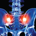
(Reuters Health)—Many doctors who order computed tomography (CT) or magnetic resonance imaging (MRI) scans for patients with low back pain do so fearing that patients will be upset if they do not get imaging and because there is too little time to explain the risks and benefits of the tests, a new study found.

The Choosing Wisely campaign, launched by the American Board of Internal Medicine in 2012, recommends against imaging scans for low back pain unless there are specific “red flags.” But a study earlier this year found that almost a third of lumbosacral MRI scans in the Department of Veterans Affairs (VA) were “inappropriate,” the authors note.

ad goes here:advert-1
ADVERTISEMENT
SCROLL TO CONTINUE

“Overuse of diagnostic tests is a common problem in healthcare as a whole, and affects both the VA and private-sector settings,” says coauthor Dr. Erika D. Sears of the VA Center for Clinical Management Research in Ann Arbor, Mich. “Low back pain is often highlighted because it is a common condition where overuse of imaging or treatments can consume a high level of resources.”

Sears and colleagues surveyed 579 VA physicians, nurse practitioners and physician assistants, including demographic questions and a hypothetical scenario in which a 45-year-old woman with nonspecific low back pain requested a CT scan or MRI.

ad goes here:advert-2
ADVERTISEMENT
SCROLL TO CONTINUE

Only 3% of clinicians thought the patient would benefit from having a CT or MRI scan, and more than three-quarters said they would worry such a scan would lead to more unnecessary tests or procedures.

But just as many also felt they would not be able to refer the woman to a specialist without obtaining imaging first, and more than half worried she would be upset if she didn’t get the imaging she wanted.

About 15% of clinicians thought it would be difficult for them to follow the recommendation against imaging, but 63% believed it would be difficult for most patients to accept it, as reported in JAMA Internal Medicine on Oct. 17.1

“You see the headlines of major sports stars getting an MRI tomorrow, so some high school athlete or college athlete or recreational athlete thinks if Derek Jeter got one why don’t I,” says J. C. Andersen, chair of the department of Health Sciences and Human Performance at The University of Tampa, Florida, who was not part of the new study.

“Tony Romo of the Cowboys got an MRI, but they’ve got lots of resources that the rest of us don’t have, and it may or may not have helped him get any better,” Andersen says.
Page: 1 2 | Single Page
Share: 

Filed under:ConditionsSoft Tissue Pain

Related Articles

    Imaging in Ankylosing Spondylitis

    April 1, 2015

    MRI inflammation, fat and new bone formation in the sacroiliac joints, spine in patients with AS

    Envision Arthritis Pathology

    April 1, 2008

    MRI advances in RA and OA

    Imaging of Axial Psoriatic Arthritis

    May 9, 2022

    The axial phenotype of psoriatic arthritis (axPsA) is an excellent example of a major controversy in rheumatology that has become the focus of attention because of the emergence of new therapies with different mechanisms of action for alleviating joint inflammation. It was first described in 1961 but, until recently, it has largely remained under the…

    A Primer on Imaging in Myositis

    November 9, 2022

    In medicine, as in advertising, pictures can be worth a thousand words. From arthritis to vasculitis, imaging studies have been variably employed to aid in the diagnosis, treatment, risk stratification and prognostication of patients with rheumatic and musculoskeletal disorders. The same holds true with the idiopathic inflammatory myopathies (IIM), in which the clinical utility is…

  • About Us
  • Meet the Editors
  • Issue Archives
  • Contribute
  • Advertise
  • Contact Us
  • Copyright © 2025 by John Wiley & Sons, Inc. All rights reserved, including rights for text and data mining and training of artificial technologies or similar technologies. ISSN 1931-3268 (print). ISSN 1931-3209 (online).
  • DEI Statement
  • Privacy Policy
  • Terms of Use
  • Cookie Preferences