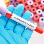
Classification criteria include the clinical presentation and the stable presence of anti-β2GPI antibodies. The lupus anticoagulant test, also part of the criteria, yields positive results with high titers of certain antibodies, most notably anti-β2GPI antibodies and the anti-phosphatidylserine/prothrombin (anti-PS/PT) antibodies.
“Clinically available testing routinely informs the clinician about three anti-β2GPI isotypes: IgG, IgM and IgA. Of these, anti-β2GPI IgG is thought to be most relevant to [the] diagnosis and pathogenesis [of APS],” say the authors.
Meanwhile, the only treatment approach to reduce thrombosis in APS is systemic anticoagulation, usually with vitamin K antagonists, such as warfarin. However, up to one in five patients experience a macrovascular thrombotic event while on systemic anticoagulation. Additionally, anticoagulants fail to protect the microvasculature, causing organ deterioration in some patients.
“There is an unmet need for safe and effective therapies that can combat the inflammatory, anticoagulant-resistant manifestations of APS,” say the authors.
“Given the diverse and likely synergistic mechanisms downstream of APS autoantibodies, we will make the case that eliminating pathogenic autoantibodies is the only strategy likely to be fully effective in treating and eventually curing patients living with APS,” they add.
Patient Case Study
The case study follows a 37-year-old woman looking for guidance on how to manage her APS. Because she had a lower extremity deep vein thrombosis at age 19—linked to initiation of an oral contraceptive—and required six months of anticoagulation therapy, the patient has since avoided estrogen-containing medications.
At age 31, she saw a rheumatologist after developing inflammatory joint pain, particularly in her hands and wrists. Laboratory testing at that time revealed a mild elevation in the erythrocyte sedimentation rate (32 mm/hr), a positive anti-cardiolipin IgG antibody result of 92 IgG phospholipid (GPL) units and a positive lupus anticoagulant test result. Anti-β2GPI antibody testing did not take place and other lupus-
relevant laboratory tests came back negative. Although prescribed hydroxychloroquine, anticoagulation was not recommended, probably due to the thrombotic event 12 years prior. Several months later, testing for aPL antibody remained similarly positive.
At age 32, the patient presented with acute abdominal pain arising during recovery from an influenza-like illness. A computed tomography scan showed colonic ischemia requiring right colon resection, and colon pathology revealed minute organizing fibrin thrombi in superficial submucosal venules. The patient required temporary dialysis for acute kidney injury and a kidney biopsy indicated acute thrombotic microangiopathy. Persistent anti-cardiolipin IgG antibody and lupus anticoagulant positivity were confirmed with laboratory testing, which also showed positive anti-β2GPI IgG and IgA isotype antibodies. In‑patient treatment comprised unfractionated heparin, high-dose glucocorticoids and plasmapheresis.

