Video: Every Case Tells a Story| Webinar: ACR/CHEST ILD Guidelines in Practice

An official publication of the ACR and the ARP serving rheumatologists and rheumatology professionals

  • Conditions
    • Axial Spondyloarthritis
    • Gout and Crystalline Arthritis
    • Myositis
    • Osteoarthritis and Bone Disorders
    • Pain Syndromes
    • Pediatric Conditions
    • Psoriatic Arthritis
    • Rheumatoid Arthritis
    • Sjögren’s Disease
    • Systemic Lupus Erythematosus
    • Systemic Sclerosis
    • Vasculitis
    • Other Rheumatic Conditions
  • FocusRheum
    • ANCA-Associated Vasculitis
    • Axial Spondyloarthritis
    • Gout
    • Psoriatic Arthritis
    • Rheumatoid Arthritis
    • Systemic Lupus Erythematosus
  • Guidance
    • Clinical Criteria/Guidelines
    • Ethics
    • Legal Updates
    • Legislation & Advocacy
    • Meeting Reports
      • ACR Convergence
      • Other ACR meetings
      • EULAR/Other
    • Research Rheum
  • Drug Updates
    • Analgesics
    • Biologics/DMARDs
  • Practice Support
    • Billing/Coding
    • EMRs
    • Facility
    • Insurance
    • QA/QI
    • Technology
    • Workforce
  • Opinion
    • Patient Perspective
    • Profiles
    • Rheuminations
      • Video
    • Speak Out Rheum
  • Career
    • ACR ExamRheum
    • Awards
    • Career Development
  • ACR
    • ACR Home
    • ACR Convergence
    • ACR Guidelines
    • Journals
      • ACR Open Rheumatology
      • Arthritis & Rheumatology
      • Arthritis Care & Research
    • From the College
    • Events/CME
    • President’s Perspective
  • Search

Envision Arthritis Pathology

Philip G. Conaghan, MB BS, PhD  |  Issue: April 2008  |  April 1, 2008

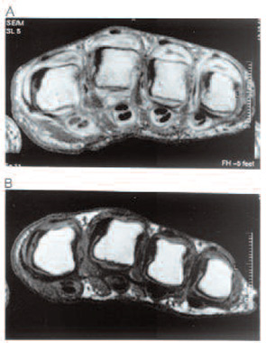
Synovitis and Tenosynovitis: Contrast-enhanced MRI shows inflammation and damage simultaneously. (See Figure 1, right.) Studies in the wrist and metacarpophalangeal joints have demonstrated that the volume of synovitis can predict subsequent erosion progression. MRI studies have demonstrated a clear relationship between inflammation and progression of bony damage at the individual joint level.3 Longitudinal studies of MRI tenosynovitis at the wrist have also demonstrated predictive validity for subsequent tendon rupture.

Bone Edema: Bone edema is one of the most interesting features to emerge from MRI studies. Radiologists first reported this feature in the late 1980s. The appearance can refer to a range of pathological processes, and is descriptive for high signal lesions in bone on fat-suppressed or STIR images. In RA, bone edema refers to a pathological process that spans the continuum from activity to damage. Early studies demonstrated that bone edema is proportional to the adjacent degree of synovitis; recent studies have confirmed that areas of edema represent an inflammatory cell infiltrate or osteitis.4 If this osteitis persists, then trabecular bone loss occurs and an erosion is formed. However, effective treatment can reduce the early bone edema lesion and thereby stop the erosion formation.
ad goes here:advert-1
ADVERTISEMENT
SCROLL TO CONTINUE
FIGURE 1: Axial sequences of rheumatoid arthritis MCP joints from the same subject before (A) and after (B) treatment with an anti-TNF therapy. (Both images are post-contrast.)
Source: Arthritis Rheum. 2002;46(7):1971-1972.
FIGURE 1: Axial sequences of rheumatoid arthritis MCP joints from the same subject before (A) and after (B) treatment with an anti-TNF therapy. (Both images are post-contrast.)

MRI in RA Research

MRI is ideal for studies on pathogenesis and therapeutic outcome because of its increased sensitivity for bony damage and ability to simultaneously visualize inflammation. Studies have demonstrated that imaging one wrist and four metacarpophalangeal (MCP) joints in a single hand is more sensitive (by a factor of 2.5) than bilateral hand and feet radiographs in detecting erosion progression over a 12-month period.5 Studying synovitis in RA trials generally requires intravenous contrast use and is often limited to studying either a single wrist or four MCP joints.

Quantification of MRI-detected RA pathology is continually improving. There are detailed volumetric methods for measuring erosion size and synovitis volume. Furthermore, dynamic contrast enhanced MRI can be used to assess synovitis, although generally it is limited to single site studies. One of the most commonly used MRI scoring systems is the RA MRI score developed by OMERACT, which is a semi-quantitative score evaluating synovitis, bone edema, and erosions. This score has been validated in a number of studies. Quantitative whole organ image analysis methods are under development and may be available within the next few years.

ad goes here:advert-2
ADVERTISEMENT
SCROLL TO CONTINUE

Page: 1 2 3 4 5 6 | Single Page
Share: 

Filed under:ConditionsOsteoarthritis and Bone Disorders Tagged with:Magnetic resonance imaging (MRI)MRIosteoarthritis (OA)PatheogenesisRheumatoid Arthritis (RA)

Related Articles

    Basics of Biologic Joint Reconstruction

    April 6, 2012

    For young patients especially, this can delay knee replacement and provide better outcomes.

    Imaging in Ankylosing Spondylitis

    April 1, 2015

    MRI inflammation, fat and new bone formation in the sacroiliac joints, spine in patients with AS

    Post-Traumatic Osteoarthritis: Managing OA That Develops After Joint Injuries & Reconstructive Surgery

    November 28, 2018

    CHICAGO—Joint trauma is one of many potential drivers of osteoarthritis disease activity and structural progression. In Post-Traumatic OA: Pathogenesis, Clinical Evolution and Management, a session at the 2018 ACR/ARHP Annual Meeting, experts discussed the effects of sports and other injuries on even young patients’ joints. Post-traumatic osteoarthritis (OA) may account for 12% of hip, knee…

    New Therapeutics for Osteoarthritis May Be in Sight

    April 1, 2015

    Overview of OA pathogenesis, recent discoveries suggest new treatment strategies are possible

  • About Us
  • Meet the Editors
  • Issue Archives
  • Contribute
  • Advertise
  • Contact Us
  • Copyright © 2025 by John Wiley & Sons, Inc. All rights reserved, including rights for text and data mining and training of artificial technologies or similar technologies. ISSN 1931-3268 (print). ISSN 1931-3209 (online).
  • DEI Statement
  • Privacy Policy
  • Terms of Use
  • Cookie Preferences