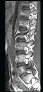
This sagittal T1 weighted MRI image of the lumbar spine shows spondylosis and bone marrowedema within the L5 pedicle extending into the articular facets and lamina.
Imae Credit: Living Art Enterprises/sciencesource.com
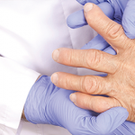
ROME, Italy—The explosion of imaging technology has made it more important than ever to establish a standardized way in which imaging can and should be used in clinical practice, an expert said in a session at EULAR 2015, the annual congress of the European League Against Rheumatism (EULAR).
Marie-Antonietta d’Agostino, MD, PhD, professor of rheumatology at the University of Paris, said clinicians now face a staggering array of choices and need guidance. “If you look at the number of imaging techniques that we can use today for our patient management, it’s completely crazy,” she said.