Video: Every Case Tells a Story| Webinar: ACR/CHEST ILD Guidelines in Practice

An official publication of the ACR and the ARP serving rheumatologists and rheumatology professionals

  • Conditions
    • Axial Spondyloarthritis
    • Gout and Crystalline Arthritis
    • Myositis
    • Osteoarthritis and Bone Disorders
    • Pain Syndromes
    • Pediatric Conditions
    • Psoriatic Arthritis
    • Rheumatoid Arthritis
    • Sjögren’s Disease
    • Systemic Lupus Erythematosus
    • Systemic Sclerosis
    • Vasculitis
    • Other Rheumatic Conditions
  • FocusRheum
    • ANCA-Associated Vasculitis
    • Axial Spondyloarthritis
    • Gout
    • Psoriatic Arthritis
    • Rheumatoid Arthritis
    • Systemic Lupus Erythematosus
  • Guidance
    • Clinical Criteria/Guidelines
    • Ethics
    • Legal Updates
    • Legislation & Advocacy
    • Meeting Reports
      • ACR Convergence
      • Other ACR meetings
      • EULAR/Other
    • Research Rheum
  • Drug Updates
    • Analgesics
    • Biologics/DMARDs
  • Practice Support
    • Billing/Coding
    • EMRs
    • Facility
    • Insurance
    • QA/QI
    • Technology
    • Workforce
  • Opinion
    • Patient Perspective
    • Profiles
    • Rheuminations
      • Video
    • Speak Out Rheum
  • Career
    • ACR ExamRheum
    • Awards
    • Career Development
  • ACR
    • ACR Home
    • ACR Convergence
    • ACR Guidelines
    • Journals
      • ACR Open Rheumatology
      • Arthritis & Rheumatology
      • Arthritis Care & Research
    • From the College
    • Events/CME
    • President’s Perspective
  • Search

Existing CT Scans as Good as DXA for Assessing Hip Fracture Risk

Scott Baltic  |  May 8, 2018

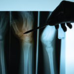
NEW YORK (Reuters Health)—A “biomechanical” analysis of a previously taken pelvic or abdominal computed tomography (CT) scan is at least as accurate in assessing an individual’s hip fracture risk as a dual-energy X-ray absorptiometry (DXA) scan, according to new research.

This accuracy of the hip bone mineral density (BMD) T-score as measured by the biomechanical CT (BCT) analysis was consistent for all fracture-risk metrics and both sexes, researchers found in a retrospective case-cohort study of nearly 4,000 participants.

ad goes here:advert-1
ADVERTISEMENT
SCROLL TO CONTINUE

Further, combining the classifications of fragile femoral bone strength and hip osteoporosis from BCT data resulted in higher sensitivity than the standard hip/spine osteoporosis classification by DXA, with comparable specificity.

In people who are already having a CT done for some other reason, particularly those aged 65 or older, “we can now screen them for osteoporosis simultaneously, sparing them the need for a separate DXA scan,” lead author Dr. Annette Adams of Kaiser Permanente Southern California tells Reuters Health by email. “Additionally, by screening the CT scans, we may pick up some people with osteoporosis or fragile bone strength who wouldn’t otherwise be screened by DXA.”

ad goes here:advert-2
ADVERTISEMENT
SCROLL TO CONTINUE

“This is significant,” she adds, because if an individual is identified as having osteoporosis, they can receive care to reduce the risk of a fracture. Such care could include medication, taking calcium and vitamin D, regular exercise and avoiding excessive tobacco and alcohol.

“When you save an older person from a fracture, you just may be saving their life,” Dr. Adams says.

The findings were published online April 17 in the Journal of Bone and Mineral Research.1

The researchers used data on more than 111,000 Kaiser Permanente patients who were 65 or older and had had a DXA scan within three years of a CT scan and no hip fracture before either scan. From this population, they matched 1,959 patients who later experienced a first non-traumatic hip fracture with 1,979 who did not.

BCT analyses of all participants’ existing CT scans were performed at a single lab that was blinded as to hip fracture status, clinical factors and DXA date.

Patients who fractured a hip were older, had lower BMD and lower body mass index and included more whites and fewer Asians, blacks and Hispanics.

Combining the classifications of fragile femoral bone strength and hip osteoporosis from BCT data resulted in higher sensitivity for predicting hip fracture than the standard hip/spine osteoporosis classification by DXA (0.66 vs. 0.59 for women; 0.56 vs. 0.48 for men), with comparable specificity (0.66 vs. 0.67 and 0.76 vs. 0.78, respectively).

Page: 1 2 3 | Single Page
Share: 

Filed under:ConditionsOsteoarthritis and Bone Disorders Tagged with:computed tomographyCT scanOsteoporosisscan

Related Articles

    Unexpected Benefits of Bisphosphonates after Hip Fracture

    February 3, 2012

    Recent trials show this bisphosphonates can reduce subsequent hip fractures and mortality, while remaining cost effective.

    Build Up Bone

    June 1, 2007

    Current management of osteoporosis

    Prevent Osteoporotic Fractures with a Fracture Liaison Service

    May 18, 2019

    Imagine leaving the hospital after suffering a heart attack without being treated for hypertension or being started on a beta blocker. What would we think of the hospital where patients are never educated about the relationship between treating hypertension and reducing myocardial infarction and stroke risk? Unfortunately, this happens every day with osteoporosis and fractures…

    Osteoporosis Experts Discuss Bisphosphonate Holidays

    November 24, 2020

    ACR CONVERGENCE 2020—Bisphosphonates are an important treatment for millions of older Americans with osteoporosis because the drugs inhibit osteoclastic bone resorption to reduce the risk of painful, debilitating fractures.1 More than 20 years ago, data emerged that bisphosphonates have a long terminal half-life.2 So after years of therapy, could some patients take a drug holiday?…

  • About Us
  • Meet the Editors
  • Issue Archives
  • Contribute
  • Advertise
  • Contact Us
  • Copyright © 2025 by John Wiley & Sons, Inc. All rights reserved, including rights for text and data mining and training of artificial technologies or similar technologies. ISSN 1931-3268 (print). ISSN 1931-3209 (online).
  • DEI Statement
  • Privacy Policy
  • Terms of Use
  • Cookie Preferences