Video: Every Case Tells a Story| Webinar: ACR/CHEST ILD Guidelines in Practice

An official publication of the ACR and the ARP serving rheumatologists and rheumatology professionals

  • Conditions
    • Axial Spondyloarthritis
    • Gout and Crystalline Arthritis
    • Myositis
    • Osteoarthritis and Bone Disorders
    • Pain Syndromes
    • Pediatric Conditions
    • Psoriatic Arthritis
    • Rheumatoid Arthritis
    • Sjögren’s Disease
    • Systemic Lupus Erythematosus
    • Systemic Sclerosis
    • Vasculitis
    • Other Rheumatic Conditions
  • FocusRheum
    • ANCA-Associated Vasculitis
    • Axial Spondyloarthritis
    • Gout
    • Psoriatic Arthritis
    • Rheumatoid Arthritis
    • Systemic Lupus Erythematosus
  • Guidance
    • Clinical Criteria/Guidelines
    • Ethics
    • Legal Updates
    • Legislation & Advocacy
    • Meeting Reports
      • ACR Convergence
      • Other ACR meetings
      • EULAR/Other
    • Research Rheum
  • Drug Updates
    • Analgesics
    • Biologics/DMARDs
  • Practice Support
    • Billing/Coding
    • EMRs
    • Facility
    • Insurance
    • QA/QI
    • Technology
    • Workforce
  • Opinion
    • Patient Perspective
    • Profiles
    • Rheuminations
      • Video
    • Speak Out Rheum
  • Career
    • ACR ExamRheum
    • Awards
    • Career Development
  • ACR
    • ACR Home
    • ACR Convergence
    • ACR Guidelines
    • Journals
      • ACR Open Rheumatology
      • Arthritis & Rheumatology
      • Arthritis Care & Research
    • From the College
    • Events/CME
    • President’s Perspective
  • Search

Experts Debate Merits of Peripheral MRI in Diagnosing RA

Greg Lavine  |  Issue: November 2008  |  November 1, 2008

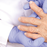
Magnetic resonance imaging (MRI) procedures have opened new windows into examining joints, but questions remain about the added value of these images in routine rheumatology practice. A session at the ACR/ARHP Annual Scientific Meeting in Boston last November focused on how results obtained from peripheral, or extremity, MRI devices should guide early intervention in rheumatoid arthritis (RA) when considering the use of biologics.

“You can’t help but be struck by the accuracy, by the beautiful nature of the pictures we have all come to recognize,” said Arthur Kavanaugh, MD, professor of medicine at the University of California, San Diego School of Medicine in La Jolla, Calif., referring to MRI-produced images.

ad goes here:advert-1
ADVERTISEMENT
SCROLL TO CONTINUE

The most established technique for rheumatologists to visualize joints remains the traditional x-ray, which is still the gold standard for diagnosing and staging RA, said Dr. Kavanaugh, who moderated the session. As the option for using MRI has started to enter the field of rheumatology, most experts agree that the new images show much more detail than x-rays.

Good—but Good Enough?

The question facing rheumatologists is whether this added level of sensitivity moves diagnosis or treatment of RA patients forward enough. Dr. Kavanaugh wondered whether these new details provide insights that could not be found using the existing array of available tools.

ad goes here:advert-2
ADVERTISEMENT
SCROLL TO CONTINUE

One drawback for MRI images, including peripheral and full-body devices, is the lack of an accepted scoring system to assess what is being seen. For x-rays as well as CT scans, there are established and widely used scoring systems to determine the extent of bone erosion and other related RA problems.

Traditional x-rays are often preferred when looking into potentially damaged joints because the procedure is widely available, easy to perform, well tolerated by patients, and relatively inexpensive. Full-body MRI procedures are less available and more expensive, and some patients feel uncomfortable lying in an enclosed space.

Peripheral MRIs offer potential solutions to some of these challenges. The portable nature of the technique means it can be placed on the target joints, which many patients prefer to having to lay in a full MRI machine. The peripheral MRI machines are priced so that individual practices could purchase them, making them potentially widely available.

But experts, including an ACR Extremity MRI Task Force that examined the merits of these portable devices, still have questions.

While the full MRIs produce high magnetic fields, many as high as 3 Teslas, the peripheral versions produce low fields, some as small as 0.2 Teslas.

Page: 1 2 3 | Single Page
Share: 

Filed under:Clinical Criteria/GuidelinesConditionsPractice SupportRheumatoid Arthritis Tagged with:MRIPeripheral MRIPractice ManagementRARheumatoid arthritis

Related Articles
    Rheumatoid Arthritis Research Provides New Insights on Risk Factors, Identification Tools, Intervention

    Rheumatoid Arthritis Research Provides New Insights on Risk Factors, Identification Tools, Intervention

    October 11, 2016

    Established wisdom holds that patients with rheumatoid arthritis (RA) will fare better if their disease is diagnosed as early as possible, and treatments with disease-modifying drugs are started before inflammation can do more damage to joints and tissue. Usually, early diagnosis means spotting the clinical signs of disease, but new research tells us more about…

    Imaging in Ankylosing Spondylitis

    April 1, 2015

    MRI inflammation, fat and new bone formation in the sacroiliac joints, spine in patients with AS

    Envision Arthritis Pathology

    April 1, 2008

    MRI advances in RA and OA

    A Duet of Bone and the Immune System

    July 12, 2011

    Examining emerging perspectives in osteoimmunology

  • About Us
  • Meet the Editors
  • Issue Archives
  • Contribute
  • Advertise
  • Contact Us
  • Copyright © 2025 by John Wiley & Sons, Inc. All rights reserved, including rights for text and data mining and training of artificial technologies or similar technologies. ISSN 1931-3268 (print). ISSN 1931-3209 (online).
  • DEI Statement
  • Privacy Policy
  • Terms of Use
  • Cookie Preferences