Video: Every Case Tells a Story| Webinar: ACR/CHEST ILD Guidelines in Practice

An official publication of the ACR and the ARP serving rheumatologists and rheumatology professionals

  • Conditions
    • Axial Spondyloarthritis
    • Gout and Crystalline Arthritis
    • Myositis
    • Osteoarthritis and Bone Disorders
    • Pain Syndromes
    • Pediatric Conditions
    • Psoriatic Arthritis
    • Rheumatoid Arthritis
    • Sjögren’s Disease
    • Systemic Lupus Erythematosus
    • Systemic Sclerosis
    • Vasculitis
    • Other Rheumatic Conditions
  • FocusRheum
    • ANCA-Associated Vasculitis
    • Axial Spondyloarthritis
    • Gout
    • Psoriatic Arthritis
    • Rheumatoid Arthritis
    • Systemic Lupus Erythematosus
  • Guidance
    • Clinical Criteria/Guidelines
    • Ethics
    • Legal Updates
    • Legislation & Advocacy
    • Meeting Reports
      • ACR Convergence
      • Other ACR meetings
      • EULAR/Other
    • Research Rheum
  • Drug Updates
    • Analgesics
    • Biologics/DMARDs
  • Practice Support
    • Billing/Coding
    • EMRs
    • Facility
    • Insurance
    • QA/QI
    • Technology
    • Workforce
  • Opinion
    • Patient Perspective
    • Profiles
    • Rheuminations
      • Video
    • Speak Out Rheum
  • Career
    • ACR ExamRheum
    • Awards
    • Career Development
  • ACR
    • ACR Home
    • ACR Convergence
    • ACR Guidelines
    • Journals
      • ACR Open Rheumatology
      • Arthritis & Rheumatology
      • Arthritis Care & Research
    • From the College
    • Events/CME
    • President’s Perspective
  • Search

Fellows’ Forum Case Report: Progressive Multifocal Leukoencephalopathy and RA

Eugene Jalbert, DO, MBA, Priyanka Murali, DO, Rakhee Shah, DO, Robert DiGiovanni, DO, FACOI, FACR, & Rubaiya Mallay, DO, FACOI, FACR  |  Issue: September 2017  |  September 18, 2017

A brain CT and an MRI (see Figure 1) were not consistent with a stroke. He was diagnosed with possible posterior reversible encephalopathy syndrome (PRES) and was discharged to a rehabilitation facility for physical and occupational therapy.

ad goes here:advert-1
ADVERTISEMENT
SCROLL TO CONTINUE
An MRI of the brain performed without contrast on June 25, 2016, showed extensive and nonspecific signal abnormalities in the cortical ribbon and subcortical white matter of the right frontal lobe. There is minimal associated parenchymal atrophy in this region without evident edema. No pathologic enhancement, diffusion restriction nor hemosiderin deposition were associated with such signal abnormality.

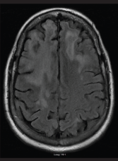
FIGURE 1: An MRI of the brain performed without contrast on June 25, 2016, showed extensive and nonspecific signal abnormalities in the cortical ribbon and subcortical white matter of the right frontal lobe. There is minimal associated parenchymal atrophy in this region without evident edema. No pathologic enhancement, diffusion restriction nor hemosiderin deposition were associated with such signal abnormality.

An MRI of the brain performed without contrast on July 7, 2016, showed persistent abnormalities in the frontal lobes, which in the right extended into the anterior adjacent parietal lobe. The abnormalities were greater in the right lobe than in the left. Findings in the occipital lobes did not have convincing evidence of restricted diffusion or hemorrhage.

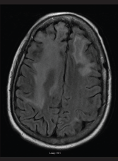
FIGURE 2: An MRI of the brain performed without contrast on July 7, 2016, showed persistent abnormalities in the frontal lobes, which in the right extended into the anterior adjacent parietal lobe. The abnormalities were greater in the right lobe than in the left. Findings in the occipital lobes did not have convincing evidence of restricted diffusion or hemorrhage.

He continued to deteriorate neurologically, which prompted a second neurology consultation. A repeat MRI (see Figure 2) and lumbar puncture were done (see Table 2). Electroencephalogram (EEG) was conducted and was consistent with mild encephalopathy. Laboratory data (see Table 2) supported a hypogammaglobulinemia with low IgG and IgM. IgA was within the normal range. CD4 and CD8 values were low. Cerebral spinal fluid (CSF) results were fairly bland, with the exception of a mild increase in RBCs. Further CSF studies, which were normal, included VDRL, EBV and oligoclonal bands. Cytology of the CSF demonstrated mature lymphocytes without malignant cells present. The CSF did demonstrate a significant elevation in myelin basic protein with a positive PCR for the JC virus.

Our patient was diagnosed with PML based on the above findings. Unfortunately, there are no FDA-approved treatment modalities for this disease process. We discussed potential experimental treatment options with the patient, but we agreed on palliative care alone.

ad goes here:advert-2
ADVERTISEMENT
SCROLL TO CONTINUE

Aggressive physical and occupational therapy were continued, and the patient was discharged home with hospice. He passed away a few months after the initial diagnosis was made.

Discussion

Progressive multifocal leukoencephalopathy (PML) is a rare, fatal, demyelinating disease of the CNS caused by lytic replication of the John Cunningham (JC) virus. The JC virus is ubiquitous in the human body and has an estimated prevalence of 40–70% in the general population.1 The JC virus is usually kept in check by the healthy immune system. However, in the right setting, the virus will invade the CNS and cause an untreatable disease state. The transformation from benign bystander to deadly disease is facilitated by an immunocompromised host, either by infection with the human immunodeficiency virus (HIV) or by immunosuppressive drugs.

Page: 1 2 3 4 | Single Page
Share: 

Filed under:ConditionsResearch RheumRheumatoid Arthritis Tagged with:ClinicalDiagnosisfellowFellowsFellows Forumoutcomeprogressive multifocal leukoencephalopathyResearchRheumatoid arthritisrheumatologistrheumatology

Related Articles

    Drug Updates

    July 1, 2009

    Information on New Approvals and Medication Safety

    Is B a Key to Autoimmune Therapy?: B Cell–targeted Therapies in Autoimmune Disease

    January 1, 2010

    A perspective on B cell–targeted therapies in autoimmune disease

    Potential Therapeutic Target for SpA

    January 23, 2018

    Innate lymphoid cells (ILCs) in the joints of patients may drive the pathology of spondyloarthritis through the production of granulocyte colony-stimulating factor (GM-CSF). A recent study found that both GM-CSF-producing CD4 T cells and the GM-CSF+ Th17 cells expressed high levels of GPR65—indicating that both GM-CSF and GPR65 may be therapeutic targets for spondyloarthritis…

    Drug Updates

    January 1, 2009

    Information on safety, labeling changes, and pharmaceutical research

  • About Us
  • Meet the Editors
  • Issue Archives
  • Contribute
  • Advertise
  • Contact Us
  • Copyright © 2025 by John Wiley & Sons, Inc. All rights reserved, including rights for text and data mining and training of artificial technologies or similar technologies. ISSN 1931-3268 (print). ISSN 1931-3209 (online).
  • DEI Statement
  • Privacy Policy
  • Terms of Use
  • Cookie Preferences