Video: Every Case Tells a Story| Webinar: ACR/CHEST ILD Guidelines in Practice

An official publication of the ACR and the ARP serving rheumatologists and rheumatology professionals

  • Conditions
    • Axial Spondyloarthritis
    • Gout and Crystalline Arthritis
    • Myositis
    • Osteoarthritis and Bone Disorders
    • Pain Syndromes
    • Pediatric Conditions
    • Psoriatic Arthritis
    • Rheumatoid Arthritis
    • Sjögren’s Disease
    • Systemic Lupus Erythematosus
    • Systemic Sclerosis
    • Vasculitis
    • Other Rheumatic Conditions
  • FocusRheum
    • ANCA-Associated Vasculitis
    • Axial Spondyloarthritis
    • Gout
    • Psoriatic Arthritis
    • Rheumatoid Arthritis
    • Systemic Lupus Erythematosus
  • Guidance
    • Clinical Criteria/Guidelines
    • Ethics
    • Legal Updates
    • Legislation & Advocacy
    • Meeting Reports
      • ACR Convergence
      • Other ACR meetings
      • EULAR/Other
    • Research Rheum
  • Drug Updates
    • Analgesics
    • Biologics/DMARDs
  • Practice Support
    • Billing/Coding
    • EMRs
    • Facility
    • Insurance
    • QA/QI
    • Technology
    • Workforce
  • Opinion
    • Patient Perspective
    • Profiles
    • Rheuminations
      • Video
    • Speak Out Rheum
  • Career
    • ACR ExamRheum
    • Awards
    • Career Development
  • ACR
    • ACR Home
    • ACR Convergence
    • ACR Guidelines
    • Journals
      • ACR Open Rheumatology
      • Arthritis & Rheumatology
      • Arthritis Care & Research
    • From the College
    • Events/CME
    • President’s Perspective
  • Search

Fellows’ Forum Case Report: Progressive Weakness and Debilitation with Skin Rash

Usman T. Malik, MBBS  |  Issue: December 2017  |  December 19, 2017

I pulled up her bloodwork, and the few images she had brought with her.

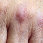
“I don’t have enough data yet,” I said, placing a hand on hers. “But judging by your history and exam,”—she stared at me—“the rash around your eyes, posterior neck, left arm, the back of your hands and feet; the way your muscles have weakened and wasted so quickly; and these hard lesions,” I pointed at the ridges of calcinosis around her joints, on her jaw, arms and pressure points, “I think you have a form of dermatomyositis.”

ad goes here:advert-1
ADVERTISEMENT
SCROLL TO CONTINUE

Her mother gripped her wheelchair. They were so quiet. I wondered for a moment if she had been a loud teenager, if she loved blasting songs from her car, maybe sang madly sometimes when she ran like the wind on a cool morning. I didn’t ask her.

Discussion

Dermatomyositis is a sinister autoimmune disease. A subset of inflammatory myopathies, the disease manifests with a spectrum of rashes most pronounced on the back of the hands, around the neck, eyes and shoulders, as well as significant muscle weakness, ranging from amyopathy to severe polymyopathy. It can start with either skin or muscle involvement and can be difficult to diagnose, especially because some of the antigen-specific antibodies that drive the inflammatory response weren’t even discovered until this past decade. More and more such antibodies are being discovered yearly, so the standard tests many community physicians rely on can produce misleading results.

ad goes here:advert-2
ADVERTISEMENT
SCROLL TO CONTINUE

A particular subset of dermatomyositis, once called dermatomyositis sine myositis and now known as clinically amyopathic dermatomyositis (CADM), can be particularly tricky to diagnose, because many patients may not have any muscle involvement at the onset of disease. The antibody most often associated with this subset, the anti-MDA5 or anti-CADM 140, is not part of the standard myositis panel and must be specifically ordered. This antibody-specific myositis can exhibit rapidly progressive interstitial lung disease, severe erosive arthritis and mechanic’s hands—cracking and thickening of the skin in the eponymous organ.

My young patient would eventually turn out to have this particular antibody.

We talked some more. I brought my attending in. We examined her together. We believed we had the correct diagnosis.

Did she look relieved? I suspect we might have given her some hope. Her eyes were brighter. When, gentle as we could, we told her we would—perhaps—not be able to restore her to prior function and that much of the damage to her joints was likely irreversible, tears slipped down her cheeks. Her chest hitched only once. I don’t know if she knew she was crying.

Page: 1 2 3 4 | Single Page
Share: 

Filed under:ConditionsMyositisOther Rheumatic Conditions Tagged with:amyopathyAutoimmune diseaseClinicaldermatomyositisDiagnosisfellowFellowsFellows Foruminflammatory myopathymuscle weaknessoutcomepatient carepolymyopathyRheumatic DiseaserheumatologistrheumatologysymptomTestTreatment

Related Articles
    Autoantibodies in Autoimmune Myopathy

    Autoantibodies in Autoimmune Myopathy

    September 18, 2017

    In recent years, scientists and clinicians have learned a great deal about autoantibodies occurring in idiopathic inflammatory myopathies (IIMs). These new discoveries have reshaped our understanding of distinct clinical pheno­types in IIMs. Scientists continue to learn more about how these auto­antibodies shape pathophysiology, diagnosis, disease monitoring, prognosis and optimum treatment. Moving forward, these autoantibodies will…

    New Tools for Myositis Diagnosis, Classification & Management

    April 15, 2019

    CHICAGO—At Hot Topics in Myositis, a session at the 2018 ACR/ARHP Annual Meeting, three experts discussed new classification criteria for idiopathic inflammatory myopathies (IIM) and offered practical primers on overlap myositis conditions and inclusion body myositis (IBM). New Myositis Classification Criteria After a 10-year development process, the new EULAR/ACR Classification Criteria for Adult and Juvenile…

    Tashatuvango / shutterstock.com

    Myositis-Specific Antibodies Identified

    January 16, 2020

    The idiopathic inflammatory myopathies (IIM) encompass eight categories: 1) dermatomyositis (DM) in adults, 2) juvenile dermatomyositis, 3) amyopathic DM, 4) cancer-associated DM, 5) polymyositis, 6) immune-mediated necrotizing myopathy, 7) inclusion body myositis, and 8) overlap myositis.1 These categories help classify the myopathies based on clinical and histologic features. The incidence of IIM is estimated at…

    Cancer-Associated Myositis: A Case Report & Review of the Literature

    Cancer-Associated Myositis: A Case Report & Review of the Literature

    February 17, 2019

    Since it was first reported in 1916, a correlation between inflammatory myopathies and cancer has been noted in several studies. Population studies have confirmed this relationship, and the phrase cancer-associated myopathy has entered the vernacular. Over the past decade, research efforts have shifted toward revealing associations between autoantibodies and clinical phenotypes. One subset of auto-antigens…

  • About Us
  • Meet the Editors
  • Issue Archives
  • Contribute
  • Advertise
  • Contact Us
  • Copyright © 2025 by John Wiley & Sons, Inc. All rights reserved, including rights for text and data mining and training of artificial technologies or similar technologies. ISSN 1931-3268 (print). ISSN 1931-3209 (online).
  • DEI Statement
  • Privacy Policy
  • Terms of Use
  • Cookie Preferences