ATLANTA—Diagnosing and treating granulomatosis with polyangiitis (GPA) and relapsing polychondritis (RP), two diseases associated with chondritis, often require patience, thoroughness and, as Philip Seo, MD, MHS, said during a session at the 2019 ACR/ARP Annual Meeting in November, time. In one case he presented during the session, perhaps a full year.

Dr. Seo
Dr. Seo is director of the Johns Hopkins Vasculitis Center, Baltimore, and the physician editor of The Rheumatologist. His talk was titled, Are Your Ears Burning? Cartilaginous Inflammation in Relapsing Polychondritis & Limited GPA. The “ears burning” part of the title illustrated part of the diagnostic dilemma: not only are GPA and RP rare, but they share symptoms of many other diseases, including manifestations that can affect the appearance and function of the ear.
Rheumatoid arthritis and spondyloarthritis are rarely associated with a finding of chondritis, according to Dr. Seo. “To me, the difference is that patients with GPA and RP generally won’t have erosions on their X-rays,” he said. Systemic lupus erythematosus is also on the list of diseases that can be associated—rarely—with chondritis, as is familial Mediterranean fever.
Being thorough to get the diagnosis right is important, because getting it wrong can ultimately be life threatening. “My philosophy is that when you’re treating a patient, you try to treat the underlying diagnosis first, rather than trying to treat the chondritis as a separate issue. If you can get the underlying disease under control, often the chondral manifestations will also go away.”
The Ears Have It
The ear is the most common site associated with chondritis and includes manifestations resembling a cauliflower ear. “This is not the only manifestation you may see,” said Dr. Seo. “In a patient with bad GPA or RP, there may be a collapse of the ear canal itself, resulting in hearing loss, because the sound can’t get through to the ear drum.”
In other patients, especially those with GPA, it is common to see a one-two punch, in which the patient develops eustachian tube inflammation followed by serous otitis media. They often complain of clogging like having water in the ear or the clogged effect after an airplane ride.
The first sign of chondral involvement in the nose is erythema and warmth in the distal septum. When that progresses, especially in GPA patients, it can lead to nasal septal perforation, often with copious amounts of crusting. “As it progresses, both GPA and RP patients can develop saddle nose deformity, where the bridge collapses,” said Dr. Seo.
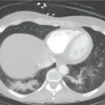
Chondritis can also affect the respiratory tract, making it too thick or too thin. “When it’s too thick, SGS [subglottic stenosis] and bronchial stenosis may result,” said Dr. Seo. “Too thin and patients develop tracheomalacia or bronchomalacia, which can lead to airway collapse.”
Some of the ocular manifestations related to RP are common, and include episcleritis/scleritis (in about 65% of cases). Dr. Seo said a broad range of ocular manifestations is associated with this diagnosis, including sicca, keratitis, optic neuritis and lid edema.
Musculoskeletal manifestations are also common. “What makes RP unique is that it can affect the sternum, so there may be involvement with the SC joints, the costochondral junctions and the sternomanubrial joints, which I think is unique for this diagnosis.”
Skin manifestations with RP are varied. They include aphthosis, purpura, livedo, urticarial, phlebitis, ulcerations and digital necrosis.
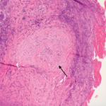
Dr. Seo said because it can be difficult to tell the difference between RP and GPA, it becomes necessary to rely on pathology to come up with a diagnosis. “For RP, that can mean seeing evidence of pleomorphic cellular infiltrate at the chondrodermal junction. Also, cartilage is gradually replaced by granulation tissue and fibrosis. For granulomatosis with polyangiitis, you may see granulomatous inflammation with multinucleated giant cells and palisading histiocytes; neutrophic, destructive angiitis; or geographic necrosis.”
Better Diagnosing May Be on the Horizon
Dr. Seo said one developing tool that may help classification in the future was presented as an abstract at the conference. (Note: Abstract No. 1719: “Clinical Phenotypes in Relapsing Polychondritis in a Prospective Cohort.”)
In the study, led by Marcela Ferrada, MD, a Lawrence Shulman Scholar at the National Institute of Arthritis and Musculoskeletal and Skin Diseases of the National Institutes of Health, Bethesda, Md., 73 patients who meet McAdams or Damiani’s diagnostic criteria for relapsing polychondritis were selected from a prospective, observational cohort. The researchers identified three phenotypes of patients with RP that differ in time to diagnosis, clinical and radiological characteristics, and complications. They were grouped as typical (14%), airway predominant (29%) or non-destructive (57%).
The researchers concluded: “Recognizing a broader spectrum of clinical patterns of disease in RP, beyond cartilaginous involvement of the ear and upper airway, may reduce diagnostic delay and facilitate development of targeted management approaches.”
As for treatment possibilities, Dr. Seo said it’s important to remember that RP is not a single diagnosis, but has a spectrum of manifestations. “So we have to make sure the punishment fits the crime—from mild to life threatening. We must also remember that many patients can be refractory to immunosuppressive therapy and may need to be cycled through several drugs to find the right ones for them to achieve remission. It’s important to remember: You will never make a diagnosis that you are not looking for.”
When Surgery Becomes Necessary
Dr. Hillel
Also presenting with Dr. Seo was his colleague and collaborator, Alexander T. Hillel, MD, an associate professor in the Department of Otolaryngology, Johns Hopkins School of Medicine. The case referred to earlier in this article was that of a patient Dr. Seo and Dr. Hillel shared. After a year, the patient was ultimately diagnosed with subglottic stenosis, which had resulted in the narrowing of his trachea.
“As an otolaryngologist, I have a bit of bias toward airway involvement when it comes to RP and GPA,” said Dr. Hillel. He stressed the importance of collaboration between rheumatologists and otolaryngologists with RA and GPA patients.
He says he avoids tracheostomies when possible, as well as open surgery. “I use endoscopic procedures. As surgeons, we must make sure to not make things worse than they have to be.”
Mike Fillon is a healthcare writer living in the Atlanta area.