Video: Every Case Tells a Story| Webinar: ACR/CHEST ILD Guidelines in Practice

An official publication of the ACR and the ARP serving rheumatologists and rheumatology professionals

  • Conditions
    • Axial Spondyloarthritis
    • Gout and Crystalline Arthritis
    • Myositis
    • Osteoarthritis and Bone Disorders
    • Pain Syndromes
    • Pediatric Conditions
    • Psoriatic Arthritis
    • Rheumatoid Arthritis
    • Sjögren’s Disease
    • Systemic Lupus Erythematosus
    • Systemic Sclerosis
    • Vasculitis
    • Other Rheumatic Conditions
  • FocusRheum
    • ANCA-Associated Vasculitis
    • Axial Spondyloarthritis
    • Gout
    • Psoriatic Arthritis
    • Rheumatoid Arthritis
    • Systemic Lupus Erythematosus
  • Guidance
    • Clinical Criteria/Guidelines
    • Ethics
    • Legal Updates
    • Legislation & Advocacy
    • Meeting Reports
      • ACR Convergence
      • Other ACR meetings
      • EULAR/Other
    • Research Rheum
  • Drug Updates
    • Analgesics
    • Biologics/DMARDs
  • Practice Support
    • Billing/Coding
    • EMRs
    • Facility
    • Insurance
    • QA/QI
    • Technology
    • Workforce
  • Opinion
    • Patient Perspective
    • Profiles
    • Rheuminations
      • Video
    • Speak Out Rheum
  • Career
    • ACR ExamRheum
    • Awards
    • Career Development
  • ACR
    • ACR Home
    • ACR Convergence
    • ACR Guidelines
    • Journals
      • ACR Open Rheumatology
      • Arthritis & Rheumatology
      • Arthritis Care & Research
    • From the College
    • Events/CME
    • President’s Perspective
  • Search

How a Nuclear Molecule Alarms the Immune System

Deena A. Abdulahad, MD, and Marc Bijl, MD  |  Issue: August 2011  |  August 1, 2011

A variety of mechanisms could explain the role of anti-dsDNA antibodies in promoting renal disease and their activity as nephrogenic or nephritogenic effector. Thus, these antibodies have the ability to bind to native glomerular structures as well as to endothelial cells. Once these immune complexes deposit in the kidney or penetrate into cells, they can act in the following ways:

  1. Modulate cell viability and induce apoptosis;
  2. Affect the proinflammatory pathways, up-regulating adhesion factors (ICAM, VCAM) in endothelial cells and IL-1, IL-8 mRNA expression in mononculear cells, and enhance IL-6 expression in glomerular mesangial cell; and
  3. Activate complement system via classical pathway leading to further tissue injury.


ad goes here:advert-1
ADVERTISEMENT
SCROLL TO CONTINUE

These effects are exerted directly by anti-dsDNA antibody via binding to Fc-receptor, which is present on the surface of many types of cell. In this situation, the receptor binds to the Fc region of antibody directly, or indirectly by DNA-containing immune complexes (via DNA antigen).16 Recently, it has been shown that these DNA-containing immune complexes also contain HMGB1, which is important for binding of these immune complexes to renal tissue and initiating renal injury in SLE patients.17

Figure 1: Inflammatory Cell.
click for large version
Figure 1: HMGB1 Release and Its Receptors. HMGB1 is released from activated inflammatory cells (e.g., macrophage). LPS, TNF, and IFN stimulate the active secretion of HMGB1 from inflammatory cells. Dying cells (apoptotic cells and/or necrotic cells) release HMGB1 as well to extracellular space passively. In turn extracellular HMGB1 acts on its target receptors (TLR-2, -4, -9, and RAGE).

Figure 2: Pathogenesis of Nephritis in SLE.

ad goes here:advert-2
ADVERTISEMENT
SCROLL TO CONTINUE
click for large version
Figure 2: Pathogenesis of Nephritis in SLE. SLE is characterized by the expression of autontibodies against nuclear components and the formation of immune complexes. Accumulated apoptotic cells are the source of autoantigens (HMGB1, nucleosome), which lead to autoantibody production and hence immune-complex formation. Circulating HMGB1 and immune complexes can promote inflammation in the kidney by several mechanisms including the interaction of HMGB1/immune complexes containing HMGB1 with renal cells via binding to RAGE, TLR-2, and TLR-4. This binding could induce apoptosis and release of proinflammatory cytokines from renal cells. Apoptotic renal cells could also accumulate and release autoantigens (nucleosomes, HMGB1). Released HMGB1 together with proinflammatory cyokines could act as chemokines attracting monocytes, macrophages, and dendritic cells. In turn, immature dendritic cells could be activated by presence of HMGB1 and migrate to lymph node where to present autoantigens and activate T–helper cells. T cells could then interact with B cells resulting in activation of B-cells and autoantibodies productions (T cell–dependent antibodies production). At the same time, B-lymphocytes could directly recognize autoantigens (HMGB1 and nucleosomes), leading further to autoantibody production through B cell receptor–TLR-9 interaction (T cell–independent antibodies production). These antibodies could bind to nuclear autoantigens and form immune complexes renal deposition; alternatively, the antibodies could bind to damaged renal structures, initiating inflammation. Finally, these deposited immune complexes/antibodies could activate the complement system via the classical pathway, amplifying renal injury.

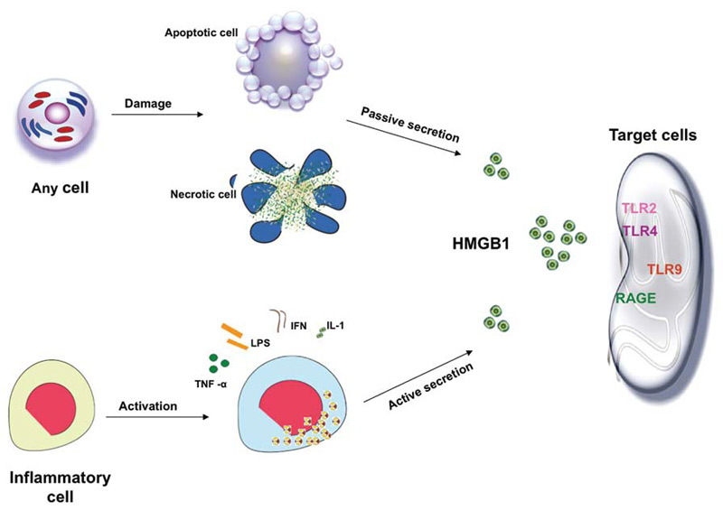
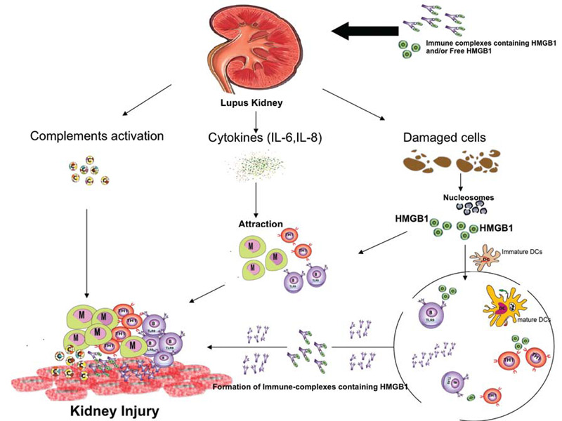
The potential role of HMGB1 in the pathogenesis of SLE is increased based on the fact that apoptotic cells accumulate in SLE and are the main source of autoantigens (nucleosomes) that may have bound HMGB1. Nucleosomes isolated form SLE sera were shown to be associated with HMGB1. Moreover, it has been shown that serum HMGB1 levels are increased in Chinese SLE patients compared with healthy controls and are correlated with disease activity and clinical parameters such as complements C3 and C4.18,19 In these studies, the investigators demonstrated that the elevation of HMGB1 levels was not associated with specific organ involvement. Also, Sato et al showed that sera of lupus nephritis patients didn’t show the tendency to be positive for HMGB1.13 However, in an IFN-accelerated murine model of SLE, administration of an anti-HMGB1 monoclonal antibody showed inhibition of proteinuria and led to kidney protection. These findings suggest that HMGB1 may play a role in the renal pathology. In contrast to previous studies on human SLE, our preliminary data show increased serum HMGB1 levels in active lupus nephritis patients.20 In addition, our data reveal a correlation of serum HMGB1 levels with proteinuria. The differences in the results of the various studies could reflect the 1) number and characteristics of patients included in the study; and 2) methods used to detect circulating HMGB1 (i.e., ELISA and Western blot).

Page: 1 2 3 4 5 6 7 | Single Page
Share: 

Filed under:ConditionsOther Rheumatic ConditionsResearch RheumSystemic Lupus Erythematosus Tagged with:HMGB1Lupus nephritisPathogenesisRenal diseaseResearch

Related Articles

    In Mouse Model, a TLR-9 Deficiency May Trigger Severe Lupus

    August 16, 2019

    Toll-like receptors play an important role in host defense. TLR-7 recognizes viral ssRNA, but also plays a role in the development of systemic lupus erythematosus (SLE). Genetic ablation of a similar receptor, TLR-9, results in opposite effects, with severe disease and kidney involvement. The mechanism of how this works remains unknown. Anna-Marie Fairhurst, PhD, from…

    Doest Rheumatoid Arthritis Take a Toll?

    November 15, 2013

    Exploring the Toll background

    A 52-Year-Old Lupus Paper Remains Important Today

    December 14, 2020

    Over 50 years ago, an article appeared in The New England Journal of Medicine: “Immunologic Factors and Clinical Activity in Systemic Lupus Erythema­tosus.”1 Written by a young postdoctoral fellow, Peter H. Schur, MD, and colleagues, the article synthesized important work in the field at the time. What follows is a discussion of the historical context…

    T Cells in Systemic Lupus Erythematosus

    August 1, 2011

    Progress toward targeted therapy

  • About Us
  • Meet the Editors
  • Issue Archives
  • Contribute
  • Advertise
  • Contact Us
  • Copyright © 2025 by John Wiley & Sons, Inc. All rights reserved, including rights for text and data mining and training of artificial technologies or similar technologies. ISSN 1931-3268 (print). ISSN 1931-3209 (online).
  • DEI Statement
  • Privacy Policy
  • Terms of Use
  • Cookie Preferences