Video: Every Case Tells a Story| Webinar: ACR/CHEST ILD Guidelines in Practice

An official publication of the ACR and the ARP serving rheumatologists and rheumatology professionals

  • Conditions
    • Axial Spondyloarthritis
    • Gout and Crystalline Arthritis
    • Myositis
    • Osteoarthritis and Bone Disorders
    • Pain Syndromes
    • Pediatric Conditions
    • Psoriatic Arthritis
    • Rheumatoid Arthritis
    • Sjögren’s Disease
    • Systemic Lupus Erythematosus
    • Systemic Sclerosis
    • Vasculitis
    • Other Rheumatic Conditions
  • FocusRheum
    • ANCA-Associated Vasculitis
    • Axial Spondyloarthritis
    • Gout
    • Psoriatic Arthritis
    • Rheumatoid Arthritis
    • Systemic Lupus Erythematosus
  • Guidance
    • Clinical Criteria/Guidelines
    • Ethics
    • Legal Updates
    • Legislation & Advocacy
    • Meeting Reports
      • ACR Convergence
      • Other ACR meetings
      • EULAR/Other
    • Research Rheum
  • Drug Updates
    • Analgesics
    • Biologics/DMARDs
  • Practice Support
    • Billing/Coding
    • EMRs
    • Facility
    • Insurance
    • QA/QI
    • Technology
    • Workforce
  • Opinion
    • Patient Perspective
    • Profiles
    • Rheuminations
      • Video
    • Speak Out Rheum
  • Career
    • ACR ExamRheum
    • Awards
    • Career Development
  • ACR
    • ACR Home
    • ACR Convergence
    • ACR Guidelines
    • Journals
      • ACR Open Rheumatology
      • Arthritis & Rheumatology
      • Arthritis Care & Research
    • From the College
    • Events/CME
    • President’s Perspective
  • Search

Inflammatory Myopathies in Children

Angela Byun Robinson, MD, and Ann M. Reed, MD  |  Issue: July 2012  |  July 10, 2012

The presence of myositis-specific autoantibodies (MSAs) is rare in children. Anti-Jo-1 antibodies are the most prevalent, found in about 2% to 5% of JDM patients, and anti-Mi-2 antibody is seen in 5% (see Table 1).11 Antibodies targeting signal recognition particles (SRP) are seen in 2% to 3% of JDM. They share the same clinical presentation as adults with an acute onset of severe myositis of both proximal and distal muscles, myocarditis, and a resultant severe disability.

ad goes here:advert-1
ADVERTISEMENT
SCROLL TO CONTINUE
click for large version
Table 1: Autoantibodies in JDM

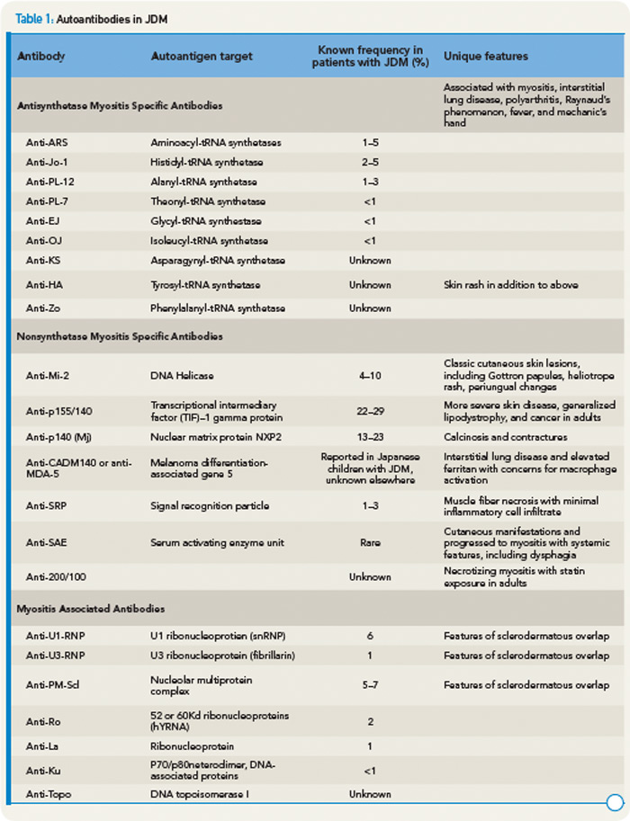
Recently, anti-p155/140 antibodies have been reported in 23% to 29% of JDM, and anti-p140 autoantibodies alone are seen in 20% to 30% of JDM and are associated with increased risk of calcinosis.12,13 Another 140kDa autoantibody, anti-MJ, is associated with severe disease with contractures and atrophy.14 While MSAs and MAAs are not always performed in children, they may be beneficial to help with prognosis and atypical clinical findings.

Recent Research Findings

It has long been appreciated that both humoral (autoantibodies and immune complexes) and cellular (T and B cells) components of the adaptive immune system contribute to the pathogenesis of JDM. However, in the last several years, there has been increasing evidence that the innate immune system, especially the upregulation of type I interferons and plasmacytoid dendritic cells may play an important role in the pathogenesis of disease.

ad goes here:advert-2
ADVERTISEMENT
SCROLL TO CONTINUE

Gene expression profiling of peripheral blood and affected tissues in JDM has revealed important insights into the molecular pathways underlying autoimmunity. The most prominent and consistent finding of these studies has been the presence of a gene signature characteristic of type I interferon (IFN) pathway activation, discovered first in DM muscle tissue and later identified in peripheral blood cells.15 The type I IFN pathway was further identified to include the cytokines/chemokines (MxA, IP-10, ITAC, MCP, interleukin 6) related to upregulation of this pathway.19

What do these studies tell us about disease mechanisms in JDM? Like many autoimmune diseases, there seems to be a genetic as well as environmental component. The genetic component appears to be multifactorial; HLA class genotype, especially HLA DQA1*0501 may help with persistence or development of maternal microchimerism.16 Cytokine polymorphisms may be involved as well. Whether there is an environmental trigger is unknown. Once the disease is initiated, there is maturation of dendritic cells leading to a type 1 interferon response and a cycle of inflammation develops with resulting damage to muscle and vascular endothelium, activation of B cells and CD4+ T cells, and overexpression of MHC class 1 on cell surfaces. It is believed that once this cycle begins, it may be halted by immunosuppressive therapy.

Therapy and Future Directions

The mortality rate of JDM was markedly reduced following the inception of corticosteroid therapy. With the use of methotrexate, the mean time of corticosteroid use fell to 10 months, compared with a historical mean time of 27 months.17 Surveys of North American pediatric rheumatologists indicate that most practitioners will treat typical JDM with corticosteroids and methotrexate, although there is great discrepancy between dosing regimens.18 Medications for refractory disease vary, but include hydroxychloroquine, intravenous immunoglobulin, rituximab, cyclophosphamide, cyclosporine A, azathioprine, tacrolimus, and mycophenolate mofetil.

Page: 1 2 3 4 5 | Single Page
Share: 

Filed under:ConditionsOther Rheumatic Conditions Tagged with:ANA titerdrugHYDROXYCHLOROQUINEinflammationinflammatory myopathyintravenous immune globulinjuvenile dermatomyositisMethotrexatepatient carePediatricsrheumatologistrituximab

Related Articles
    Oksana Kuzmina/shutterstock.comx

    Environmental Factors in Pediatric Systemic Autoimmune Diseases

    March 20, 2017

    Systemic autoimmune diseases are thought to result from immune dysregulation in genetically susceptible individuals who were exposed to environmental risk factors. Many studies have identified genetic risk factors for these diseases, but concordance rates among monozygotic twins are 25–40%, suggesting that nonheritable environmental factors play a more prominent role.1,2 Through carefully conducted epidemiologic and other…

    Updates in Juvenile Dermatomyositis: Pathogenesis & Patient Care

    July 17, 2021

    Experts provided an update on juvenile dermatomyositis, discussing patient care, treatment options and the pathogenesis of disease.

    New Tools for Myositis Diagnosis, Classification & Management

    April 15, 2019

    CHICAGO—At Hot Topics in Myositis, a session at the 2018 ACR/ARHP Annual Meeting, three experts discussed new classification criteria for idiopathic inflammatory myopathies (IIM) and offered practical primers on overlap myositis conditions and inclusion body myositis (IBM). New Myositis Classification Criteria After a 10-year development process, the new EULAR/ACR Classification Criteria for Adult and Juvenile…

    Reading RHEUM

    November 1, 2006

    Handpicked Reviews of Contemporary Literature

  • About Us
  • Meet the Editors
  • Issue Archives
  • Contribute
  • Advertise
  • Contact Us
  • Copyright © 2025 by John Wiley & Sons, Inc. All rights reserved, including rights for text and data mining and training of artificial technologies or similar technologies. ISSN 1931-3268 (print). ISSN 1931-3209 (online).
  • DEI Statement
  • Privacy Policy
  • Terms of Use
  • Cookie Preferences