Video: Every Case Tells a Story| Webinar: ACR/CHEST ILD Guidelines in Practice

An official publication of the ACR and the ARP serving rheumatologists and rheumatology professionals

  • Conditions
    • Axial Spondyloarthritis
    • Gout and Crystalline Arthritis
    • Myositis
    • Osteoarthritis and Bone Disorders
    • Pain Syndromes
    • Pediatric Conditions
    • Psoriatic Arthritis
    • Rheumatoid Arthritis
    • Sjögren’s Disease
    • Systemic Lupus Erythematosus
    • Systemic Sclerosis
    • Vasculitis
    • Other Rheumatic Conditions
  • FocusRheum
    • ANCA-Associated Vasculitis
    • Axial Spondyloarthritis
    • Gout
    • Psoriatic Arthritis
    • Rheumatoid Arthritis
    • Systemic Lupus Erythematosus
  • Guidance
    • Clinical Criteria/Guidelines
    • Ethics
    • Legal Updates
    • Legislation & Advocacy
    • Meeting Reports
      • ACR Convergence
      • Other ACR meetings
      • EULAR/Other
    • Research Rheum
  • Drug Updates
    • Analgesics
    • Biologics/DMARDs
  • Practice Support
    • Billing/Coding
    • EMRs
    • Facility
    • Insurance
    • QA/QI
    • Technology
    • Workforce
  • Opinion
    • Patient Perspective
    • Profiles
    • Rheuminations
      • Video
    • Speak Out Rheum
  • Career
    • ACR ExamRheum
    • Awards
    • Career Development
  • ACR
    • ACR Home
    • ACR Convergence
    • ACR Guidelines
    • Journals
      • ACR Open Rheumatology
      • Arthritis & Rheumatology
      • Arthritis Care & Research
    • From the College
    • Events/CME
    • President’s Perspective
  • Search

Is Ultrasound Right For YOUR Office?

Kurt Ullman  |  Issue: July 2011  |  July 12, 2011

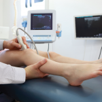
An example of a mobile, in-office ultrasound machine that can be used by rheumatology practices for diagnosis and treatment.

The uses of ultrasound (US) technology in rheumatology have been growing in importance over the last few years. Many rheumatologists are beginning to explore the possibility of adding US to their practices.

“Ultrasound is becoming the state of the art in diagnosis and treatment in the United States,” says Jonathan Samuels, MD, assistant professor of medicine in the division of rheumatology at the New York University Hospital for Joint Diseases in New York City. “It is now considered by many to be a part of our physical examination, like a rheumatologist’s other stethoscope.”

ad goes here:advert-1
ADVERTISEMENT
SCROLL TO CONTINUE

With this technology, physicians can “see” inside the joint in real time and find indications of active inflammation or fluid—indicators that are often hard to find by palpation or visual assessment alone. There are also dynamic applications that allow physicians to assess a joint during movement. This is not possible using static modalities such as magnetic resonance imaging or computed tomography scans.

Diagnostic Uses

“From the diagnostic perspective, you are able to do a lot of things as a rheumatologist that you would not feel as comfortable doing, or do as well, without it,” says Eugene Kissin, MD, associate professor of medicine at Boston University. “Sometimes we get a patient with joint pain, but nothing is found on the initial examination. It is nice to be able to see within minutes if, in fact, there is inflammation.”

ad goes here:advert-2
ADVERTISEMENT
SCROLL TO CONTINUE

US is also finding more uses in helping physicians make treatment decisions. Dr. Kissin points to the patient who seems to be doing well and may be a candidate for lowering or even stopping medications. The images from the US could show subclinical activity resulting in a reevaluation of that decision.

“In one study of people presenting with ankle pain, an ultrasound was performed after the participating rheumatologists had completed their routine examination and had formulated a care plan,” he says.1 “Most of the time, the additional information from the study caused revisions in the plan of care.”

US in Treatment

After the plans are in place, US is proving helpful during treatment. Real-time guidance of needle placement for aspiration and medication administration allows the physician to be more confident that the needle is in the exact place it needs to be.

Page: 1 2 3 4 5 | Single Page
Share: 

Filed under:Practice SupportTechnology Tagged with:diagnostic imagingpatient carerheumatologistTechnologyUltrasound

Related Articles

    How Rheumatologists Can Find, Develop Their Specialty Niche

    March 15, 2016

    Many rheumatologists chose the profession because they wanted to delve more deeply into rheumatic diseases than internal medicine calls for. “For most, this level of specialization is sufficient and worthy of a lifelong devotion and commitment,” says S. Sam Lim, MD, MPH, associate professor, Medicine and Epidemiology, Emory University School of Medicine, Atlanta. “However, some…

    Tips for Implementing Ultrasound Training in Rheumatology Fellowships

    February 13, 2020

    ATLANTA—Point-of-care ultrasound education mainly has occurred at the undergraduate level at U.S. medical schools, but rheumatology fellowship training programs are rapidly catching up and integrating it into their curricula, according to two program directors who reviewed the state of rheumatology ultrasound education, including potential barriers to its implementation, on Nov. 12 at the 2019 ACR/ARP…

    Ultrasound Training Tips & Pitfalls

    November 19, 2018

    The past 20 years have seen dramatic changes in the practice of rheumatology, ranging from bench to bedside therapeutic advances to dramatic improvements in diagnostic imaging. The results have been gratifying for our patients and attractive to internal medicine trainees making subspecialty career decisions. We are pleased to provide this article for The Rheumatologist’s wide-ranging…

    Musculoskeletal Ultrasound: A Valuable Tool for Diagnosing Rheumatic Illnesses

    Musculoskeletal Ultrasound: A Valuable Tool for Diagnosing Rheumatic Illnesses

    October 15, 2015

    Musculoskeletal (MSK) ultrasound is a valuable imaging modality for the practicing rheumatologist and provides an efficient tool with high diagnostic value in the evaluation of patients with musculoskeletal complaints. The use of MSK ultrasound has evolved in the U.S. due to the emergence of less-expensive, portable ultrasound units, which provide high-quality gray-scale and power Doppler…

  • About Us
  • Meet the Editors
  • Issue Archives
  • Contribute
  • Advertise
  • Contact Us
  • Copyright © 2025 by John Wiley & Sons, Inc. All rights reserved, including rights for text and data mining and training of artificial technologies or similar technologies. ISSN 1931-3268 (print). ISSN 1931-3209 (online).
  • DEI Statement
  • Privacy Policy
  • Terms of Use
  • Cookie Preferences