The Rheumatologist
COVID-19 News
  • Connect with us:
  • Facebook
  • Twitter
  • LinkedIn
  • YouTube
  • Feed
  • Home
  • Conditions
    • Rheumatoid Arthritis
    • SLE (Lupus)
    • Crystal Arthritis
      • Gout Resource Center
    • Spondyloarthritis
    • Osteoarthritis
    • Soft Tissue Pain
    • Scleroderma
    • Vasculitis
    • Systemic Inflammatory Syndromes
    • Guidelines
  • Resource Centers
    • Ankylosing Spondylitis Resource Center
    • Gout Resource Center
    • Rheumatoid Arthritis Resource Center
    • Systemic Lupus Erythematosus Resource Center
  • Drug Updates
    • Biologics & Biosimilars
    • DMARDs & Immunosuppressives
    • Topical Drugs
    • Analgesics
    • Safety
    • Pharma Co. News
  • Professional Topics
    • Ethics
    • Legal
    • Legislation & Advocacy
    • Career Development
      • Certification
      • Education & Training
    • Awards
    • Profiles
    • President’s Perspective
    • Rheuminations
  • Practice Management
    • Billing/Coding
    • Quality Assurance/Improvement
    • Workforce
    • Facility
    • Patient Perspective
    • Electronic Health Records
    • Apps
    • Information Technology
    • From the College
    • Multimedia
      • Audio
      • Video
  • Resources
    • Issue Archives
    • ACR Convergence
      • Systemic Lupus Erythematosus Resource Center
      • Rheumatoid Arthritis Resource Center
      • Gout Resource Center
      • Abstracts
      • Meeting Reports
      • ACR Convergence Home
    • American College of Rheumatology
    • ACR ExamRheum
    • Research Reviews
    • ACR Journals
      • Arthritis & Rheumatology
      • Arthritis Care & Research
      • ACR Open Rheumatology
    • Rheumatology Image Library
    • Treatment Guidelines
    • Rheumatology Research Foundation
    • Events
  • About Us
    • Mission/Vision
    • Meet the Authors
    • Meet the Editors
    • Contribute to The Rheumatologist
    • Subscription
    • Contact
  • Advertise
  • Search
You are here: Home / Articles / Malignant Atrophic Papulosis Is Challenging to Diagnose, Treat

Malignant Atrophic Papulosis Is Challenging to Diagnose, Treat

July 1, 2014 • By Aixa Toledo-Garcia, MD, Jessica Farrell, PharmD, & Lee Shapiro, MD

  • Tweet
  • Email
Print-Friendly Version / Save PDF

You Might Also Like
  • Giant Cell Arteritis Challenging to Diagnose, Manage
  • Cryopyrin-Associated Periodic Syndromes: Difficult to Recognize, Diagnose, Treat
  • Diagnose, Manage and Treat Giant Cell Arteritis
Explore This Issue
July 2014
State-of-the-art review of Köhlmeier-Degos disease

ad goes here:advert-1
ADVERTISEMENT
SCROLL TO CONTINUE

In December 2009, we encountered a 17-year-old male who was dying from a recently diagnosed catastrophic disease. He was previously healthy and athletic. He was a patient in the intensive care unit (ICU) with decompensated vital signs, following an exploratory laparotomy for poly­bacterial peritonitis and sepsis from gastrointestinal (GI) microperforations. He was deteriorating despite treatment with immune intravenous globulin (IVIG) and broad-spectrum antibiotics.

Prior to his presentation in the ICU, the patient had a two-year history of developing innumerable, multiplying avascular slightly depressed porcelain-like skin lesions surrounded by an erythematous rim. These were present primarily on his chest and abdomen. He was diagnosed with malignant atrophic papulosis (MAP), based on the skin biopsy findings and clinical history, in August 2009 (see Figure 1). Abdominal pain and other nonspecific GI symptoms had started six months earlier. Endoscopy was performed, which showed gastritis and an erythematous mucosa of the sigmoid colon, rectosigmoid junction and splenic flexure. Because of the severity of his pain, an exploratory laparotomy was performed, which showed multiple small 5–8 mm ulcerated lesions throughout the small bowel (see Figure 2). Further evaluation for an underlying rheumatologic disease was negative.

ad goes here:advert-2
ADVERTISEMENT
SCROLL TO CONTINUE

Figure 1: A skin biopsy and clinical history led to a diagnosis of malignant atrophic papulosis in this 17-year-old male patient.

With the patient’s health steadily deteriorating, we desperately reached out to the medical community. We heard of a similar patient who had responded to treatment with eculizumab. We were able to obtain eculizumab on a compassionate-use basis, and he had an almost immediate response. Soon after, his abdomen was closed; it had been left partially opened due to abdominal compartment syndrome. Repeat echocardiogram showed resolution of the pericardial effusion, and the chest X-ray also showed improvement.

He continued to receive eculizumab infusions every other week and self-administered subcutaneous enoxaparin daily. He improved to the point that he gained back all the weight he had lost and was exercising again. Eventually, he returned to school, work and enjoying his life with family and friends.

ad goes here:advert-3
ADVERTISEMENT
SCROLL TO CONTINUE

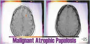
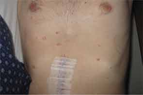
Six months after discharge, he developed gross hematuria and severe flank pain (enoxaparin was stopped). Cystoscopy demonstrated multiple lesions suggestive of MAP (see Figure 3). The dose of eculizumab was increased due to the concern that the dose reaching his bladder was subtherapeutic. The patient developed neurological symptoms, including perioral numbness and tingling sensation around his distal fingers. A magnetic resonance imaging (MRI) study of the brain was concerning for left frontal lobe vasculitis, as well as the presence of a 1 mm thick subdural hematoma. The dose of eculizumab was increased to weekly injections, and he was started on levetiracetam and aspirin.

Pages: 1 2 3 4 5 | Single Page

Filed Under: Biologics & Biosimilars, Conditions, Drug Updates, Scleroderma, SLE (Lupus), Vasculitis Tagged With: Biologics, drug, infliximab, intravenous immune globulin, Köhlmeier-Degos, lesions, malignant atrophic papulosis, patient care, rheumatologist, Scleroderma, systemic lupus erythematous, VasculitisIssue: July 2014

You Might Also Like:
  • Giant Cell Arteritis Challenging to Diagnose, Manage
  • Cryopyrin-Associated Periodic Syndromes: Difficult to Recognize, Diagnose, Treat
  • Diagnose, Manage and Treat Giant Cell Arteritis
  • Dermatology Case: History of ANCA–Associated Vasculitis, Fever, Rash

Rheumatology Research Foundation

The Foundation is the largest private funding source for rheumatology research and training in the U.S.

Learn more »

ACR Convergence

Don’t miss rheumatology’s premier scientific meeting for anyone involved in research or the delivery of rheumatologic care or services.

Visit the ACR Convergence site »

Meeting Abstracts

Browse and search abstracts from the ACR Convergence and ACR/ARP Annual Meetings going back to 2012.

Visit the Abstracts site »

The Rheumatologist newsmagazine reports on issues and trends in the management and treatment of rheumatic diseases. The Rheumatologist reaches 11,500 rheumatologists, internists, orthopedic surgeons, nurse practitioners, physician assistants, nurses, and other healthcare professionals who practice, research, or teach in the field of rheumatology.

About Us / Contact Us / Advertise / Privacy Policy / Terms of Use

  • Connect with us:
  • Facebook
  • Twitter
  • LinkedIn
  • YouTube
  • Feed

Copyright © 2006–2021 American College of Rheumatology. All rights reserved.

ISSN 1931-3268 (print)
ISSN 1931-3209 (online)

loading Cancel
Post was not sent - check your email addresses!
Email check failed, please try again
Sorry, your blog cannot share posts by email.
This site uses cookies: Find out more.