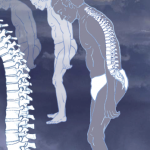
Understanding the Diagnosis of Non-Radiographic Axial Spondyloarthritis
SAN DIEGO—For many decades, the entity of non-radiographic axial spondyloarthritis (nr-axSpA) did not officially exist because patients either met or did not meet criteria for ankylosing spondylitis, which was based on radiographs. In recent years, the recognition of nr-axSpA has helped identify the cause of back pain in many patients previously without a diagnosis. However, questions remain about how to avoid under- or over-diagnosing the condition. In the session titled, Pearls and Pitfalls in Diagnosing Non-Radiographic Axial Spondyloarthritis, several speakers provided high-yield insights on this topic.
A Conceptual Approach to Diagnosis

Dr. Hwang
The first speaker was Mark Hwang, MD, assistant professor of medicine, McGovern Medical School, UTHealth, Houston, who focused on a conceptual approach to the diagnosis of nr-axSpA. Historically, the modified New York diagnostic criteria for ankylosing spondylitis required radiographic changes—specifically grade 3–4 unilateral or grade 2–4 bilateral sacroiliitis—in addition to clinical criteria for a patient to be diagnosed with the condition. In 2009, the Assessment of SpondyloArthritis international Society (ASAS) developed classification criteria that coined the terms axial spondyloarthritis (axSpA) and nr-axSpA.
If we assume that a test, such as radiographs of the sacroiliac (SI) joints, has a set sensitivity or specificity for detecting sacroiliitis, then these can be combined with the likelihood ratios associated with characteristics of the patient in order to calculate the odds of axSpA being the correct diagnosis. Thus, two patients—a 35-year-old man with five years of back pain that is associated with two hours of morning stiffness and a 45-year-old man with three months of back pain that causes 15 minutes of morning stiffness—would be regarded as having different odds of axSpA even if their imaging findings are identical.
Although this type of thinking is often characteristic of what occurs in clinical practice, Dr. Hwang noted that clinical reasoning is a constant, iterative process, one that requires an ongoing refinement of our understanding of each disease and an analysis of our clinical capabilities.
Imaging

Dr. Gensler
The second speaker in the session was Lianne Gensler, MD, professor of medicine, director, Ankylosing Spondylitis Clinic, University of California, San Francisco. She described the role of imaging in the diagnosis of nr-axSpa.
To start her talk, Dr. Gensler paraphrased the shoe company Nike: if you are thinking about pursuing SI joint imaging based on your clinical assessment, just do it. She noted that imaging is but one piece of the puzzle, but it is a helpful data point because physical exam findings can be hard to detect in patients with nr-axSpA.
Many other features of the condition are non-specific, and there are potential mimics of the disease, thus imaging can help clarify the clinical impression of a provider. In essence, just as a rheumatologist would pursue a temporal artery biopsy for a patient suspected of having giant cell arteritis, so too should they pursue imaging when features of inflammatory back pain are present.
Dr. Gensler explained that imaging in axSpA can be helpful both for diagnosis and for evaluating the effects of treatment/disease progression. Conventional radiographs remain the first-line imaging study, although they generally have poor sensitivity and specificity.
MRI of the pelvis or sacrum would be the next best test to pursue when radiographs are negative but clinical suspicion for axSpA remains high. The MRI does not require contrast and, specifically, the T1 sequence can be helpful in evaluating structural changes and the STIR/T2 fat suppressed sequence can be helpful in evaluating inflammatory changes.
In addition, low-dose computed tomography studies of the pelvis may also be helpful and are widely available.
Dr. Gensler observed that ordering and arranging for the correct imaging for patients can be complicated in terms of logistics. Further, even when the correct imaging protocols are used, there is variability among radiologists in their familiarity with axSpA and their ability to read these studies well. Ideally, a discussion between the rheumatologist and the radiologist should take place to help ensure a mutual understanding of imaging findings.
In the future, artificial intelligence may be able to assist in detecting subtle inflammatory changes and could serve as a proxy for an expert radiologist in geographic areas where such experts are not available.
Mimics & More

Dr. Ogdie-Beatty
The final speaker in the session was Alexis Ogdie-Beatty, MD, MSCE, associate professor of medicine, associate professor of epidemiology, Department of Medicine, Hospital of the University of Pennsylvania, Philadelphia. Dr. Ogdie-Beatty explained that, when evaluating for nr-axSpA, a rheumatologist should always be asking, “What else could this be?”
The list of potential mimicking conditions is long and includes mechanical back pain, fibromyalgia, degenerative disc disease, fracture, osteitis condensans ilii, diffuse idiopathic skeletal hyperostosis (DISH), sarcoidosis, crystalline arthritis and septic arthritis.
One key question with which to begin the evaluation is to ask, “Where is the pain?” The specific location of pain, as well as patient age, precipitating factors and relationship to physical activity, will often help inform if this is more or less likely to be mechanical back pain.
Dr. Ogdie-Beatty explained that inflammatory back pain is typically associated with an insidious onset, is worse at night (especially the second half of the night) and improves with activity.
In speaking about other causes of back pain, fibromyalgia is challenging because it can both mimic inflammatory back pain as well as exist as a comorbidity in up to one-third of patients with axSpA.
Osteitis condensans ilii is characterized by benign sclerosis of the ilium adjacent to the SI joints, often appearing bilaterally and with a triangular shape. The condition is more common in women than men, particularly during and after pregnancy. DISH is normally found in men over age 45 and is associated with the presence of diabetes mellitus. It may be asymptomatic and present mostly as decreased range of motion in the spine; enthesophytes are also commonly seen with this condition.
Some other mimics of inflammatory back pain, such as Paget’s disease, X-linked hypophosphatemia and alkaptonuric ochronosis (an autosomal recessive disease that results in buildup of homogentisic acid in cartilage and subsequent arthropathy) are quite rare, but worth at least being aware of.
In Sum
The session was stimulating and highly informative, which comes as a great relief to the rheumatology world that is still trying to better understand the entity of nr-axSpA.
Jason Liebowitz, MD, is an assistant professor of medicine in the Division of Rheumatology at Columbia University Vagelos College of Physicians and Surgeons, New York.
Reference
- Rudwaleit M, van der Heijde D, Landewé R, et al. The development of Assessment of SpondyloArthritis international Society classification criteria for axial spondyloarthritis (part II): Validation and final selection [published correction appears in Ann Rheum Dis. 2019 Jun;78(6):e59]. Ann Rheum Dis. 2009;68(6):777–783.