Video: Every Case Tells a Story| Webinar: ACR/CHEST ILD Guidelines in Practice

An official publication of the ACR and the ARP serving rheumatologists and rheumatology professionals

  • Conditions
    • Axial Spondyloarthritis
    • Gout and Crystalline Arthritis
    • Myositis
    • Osteoarthritis and Bone Disorders
    • Pain Syndromes
    • Pediatric Conditions
    • Psoriatic Arthritis
    • Rheumatoid Arthritis
    • Sjögren’s Disease
    • Systemic Lupus Erythematosus
    • Systemic Sclerosis
    • Vasculitis
    • Other Rheumatic Conditions
  • FocusRheum
    • ANCA-Associated Vasculitis
    • Axial Spondyloarthritis
    • Gout
    • Psoriatic Arthritis
    • Rheumatoid Arthritis
    • Systemic Lupus Erythematosus
  • Guidance
    • Clinical Criteria/Guidelines
    • Ethics
    • Legal Updates
    • Legislation & Advocacy
    • Meeting Reports
      • ACR Convergence
      • Other ACR meetings
      • EULAR/Other
    • Research Rheum
  • Drug Updates
    • Analgesics
    • Biologics/DMARDs
  • Practice Support
    • Billing/Coding
    • EMRs
    • Facility
    • Insurance
    • QA/QI
    • Technology
    • Workforce
  • Opinion
    • Patient Perspective
    • Profiles
    • Rheuminations
      • Video
    • Speak Out Rheum
  • Career
    • ACR ExamRheum
    • Awards
    • Career Development
  • ACR
    • ACR Home
    • ACR Convergence
    • ACR Guidelines
    • Journals
      • ACR Open Rheumatology
      • Arthritis & Rheumatology
      • Arthritis Care & Research
    • From the College
    • Events/CME
    • President’s Perspective
  • Search

New Study Aimed at Better Predicting Large Vessel Vasculitis Relapse

Elizabeth Hofheinz, MPH, MEd  |  Issue: September 2018  |  September 20, 2018

vitstudio / shutterstock.com

vitstudio / shutterstock.com

Imagine telling a patient “You’re in remission!” and finding out it’s not true. The last thing you want to do is get it wrong clinically and put your patient on an emotional rollercoaster.

With large vessel vasculitis (LVV) in particular, physicians struggle to be accurate, to determine if indeed the disease has gone away or just gone into hiding.

ad goes here:advert-1
ADVERTISEMENT
SCROLL TO CONTINUE

Peter Grayson, MD, MSc, is head of the Vasculitis Translational Research Program at the National Institute of Arthritis and Musculoskeletal and Skin Diseases at the National Institutes of Health (NIH) in Bethesda, Md. Dr. Grayson and colleagues published an article in the March 2018 issue of Arthritis & Rheumatology on the use of 18F-fluorodeoxyglucose (FDG) positron emission tomography (PET) as an imaging biomarker for large vessel vasculitis.1

“Like many rheumatologists,” he says, “I have had a patient with LVV who, according to the bloodwork, was in remission, but in fact was not. Determining whether the disease is still active is easier to do shortly after the initial diagnosis when things are more obvious, but it becomes more difficult over the disease course. Tests, such as the erythrocyte sedimentation rate, are not necessarily reliable to determine disease activity.

ad goes here:advert-2
ADVERTISEMENT
SCROLL TO CONTINUE

“There are fewer clinical trials in LVV [than for] forms of small vessel vasculitis, in part because there are no validated outcome measures of disease activity in LVV. “It’s hard to determine if a therapy is effective when it is simultaneously challenging to figure out if the disease is still active.”

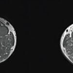
‘FDG-PET was useful in differentiating patients with clinically active LVV from controls (a sensitivity of 85% & a specificity of 83%). However, we also observed that the majority of patients with LVV in clinical remission at the time of imaging had FDG-PET scan findings that looked like ongoing vasculitis.’ —Dr. Grayson

Does Bloodwork = Guesswork?

Dr. Grayson

Dr. Grayson

“There is always the concern with LVV that medications improve the blood markers of inflammation without improving inflammation at the tissue level,” Dr. Grayson says. “In fact, when autopsies have been performed on patients with LVV who died of other causes and were thought to be in clinical remission at the time of death, ongoing active vasculitis is almost always detected in the arteries. So somehow, it appears the treatments for LVV are not getting at the underlying vascular inflammation.

“FDG-PET is traditionally used to detect metabolic activity in cancer cells,” he says. “However, it has been known for years that FDG-PET can also be used to visualize inflammation.”

Page: 1 2 3 | Single Page
Share: 

Filed under:Vasculitis Tagged with:large-vessel vasculitisrelapseRemission

Related Articles

    Get the Picture: RheumMadness 2022 PET-CT in Large Vessel Vasculitis Scouting Report

    February 14, 2022

    With both impressive sensitivity and specificity in capturing active large vessel vasculitis, a positron emission tomography (PET) scan is a potential tool for determining disease activity and predicting relapse in patients with large vessel vasculitis.

    Rheum After 5: Introducing Dr. Grayson & the Affordable Rock ‘n’ Roll Act

    January 19, 2021

    Peter Grayson, MD, MSc, a tenure track investigator at the National Institutes of Health (NIH) Intramural Research Program, Bethesda, Md., sometimes wonders how his life would have turned out if the band he was playing in during college had signed a recording contract with a major record label. Dr. Grayson has been playing the piano…

    Updates on Giant Cell Arteritis

    March 19, 2018

    SAN DIEGO—Recent research tells us more about giant cell arteritis (GCA) to help rheumatologists more accurately diagnose and effectively treat patients with this type of vasculitis. On Nov. 6 at the ACR/ARHP Annual Meeting, three experts explored the latest findings on GCA pathogenesis, diagnostic approaches, imaging modalities and growing treatment options. GCA: What’s Really Happening?…

    A Primer on Imaging in Myositis

    November 9, 2022

    In medicine, as in advertising, pictures can be worth a thousand words. From arthritis to vasculitis, imaging studies have been variably employed to aid in the diagnosis, treatment, risk stratification and prognostication of patients with rheumatic and musculoskeletal disorders. The same holds true with the idiopathic inflammatory myopathies (IIM), in which the clinical utility is…

  • About Us
  • Meet the Editors
  • Issue Archives
  • Contribute
  • Advertise
  • Contact Us
  • Copyright © 2025 by John Wiley & Sons, Inc. All rights reserved, including rights for text and data mining and training of artificial technologies or similar technologies. ISSN 1931-3268 (print). ISSN 1931-3209 (online).
  • DEI Statement
  • Privacy Policy
  • Terms of Use
  • Cookie Preferences