Fibrosing Myopathy
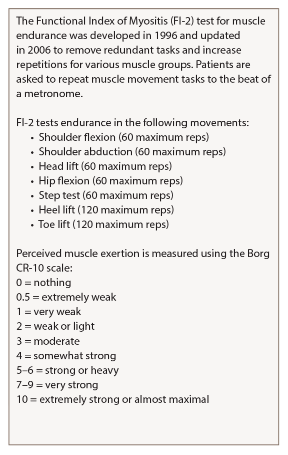
Table 2: Functional Index of Myositis
Karolinska Institute in Stockholm created a detailed guide on how to perform and score the FI-2 test that is available on the National Institute of Environmental Health Science website.
“After we found those three cases, we started to do a more systematic assessment of muscle weakness in all our scleroderma patients, and we started to see that, when we did biopsies, we started to see more of these patients,” said Dr. Paik. “So, we coined the term fibrosing myopathy, where we see fibrosis only and a lack of inflammatory infiltrates.” These patients’ fibrosis is primarily perimysial and endomysial, and thick, dense connective tissue encases the muscle fascicle, she explained.
In a 2017 study designed to further understand the clinical phenotype of fibrosing myopathy, the researchers compared these patients with those with an inflammatory myopathy, defined as inflammation and/or necrosis.16

