Video: Every Case Tells a Story| Webinar: ACR/CHEST ILD Guidelines in Practice

An official publication of the ACR and the ARP serving rheumatologists and rheumatology professionals

  • Conditions
    • Axial Spondyloarthritis
    • Gout and Crystalline Arthritis
    • Myositis
    • Osteoarthritis and Bone Disorders
    • Pain Syndromes
    • Pediatric Conditions
    • Psoriatic Arthritis
    • Rheumatoid Arthritis
    • Sjögren’s Disease
    • Systemic Lupus Erythematosus
    • Systemic Sclerosis
    • Vasculitis
    • Other Rheumatic Conditions
  • FocusRheum
    • ANCA-Associated Vasculitis
    • Axial Spondyloarthritis
    • Gout
    • Psoriatic Arthritis
    • Rheumatoid Arthritis
    • Systemic Lupus Erythematosus
  • Guidance
    • Clinical Criteria/Guidelines
    • Ethics
    • Legal Updates
    • Legislation & Advocacy
    • Meeting Reports
      • ACR Convergence
      • Other ACR meetings
      • EULAR/Other
    • Research Rheum
  • Drug Updates
    • Analgesics
    • Biologics/DMARDs
  • Practice Support
    • Billing/Coding
    • EMRs
    • Facility
    • Insurance
    • QA/QI
    • Technology
    • Workforce
  • Opinion
    • Patient Perspective
    • Profiles
    • Rheuminations
      • Video
    • Speak Out Rheum
  • Career
    • ACR ExamRheum
    • Awards
    • Career Development
  • ACR
    • ACR Home
    • ACR Convergence
    • ACR Guidelines
    • Journals
      • ACR Open Rheumatology
      • Arthritis & Rheumatology
      • Arthritis Care & Research
    • From the College
    • Events/CME
    • President’s Perspective
  • Search

Patients with Large-Vessel Abnormalities Present Diagnostic Dilemma for Rheumatologists

Eli Miloslavsky, MD  |  Issue: February 2015  |  February 1, 2015

On examination, he was afebrile, and his blood pressure was 145/85 mmHg with a heart rate of 121 beats per minute. He had easily visible veins on his shoulders, chest and abdomen but no hyperextensible joints. The remainder of the examination was unremarkable. Repeat laboratory testing revealed a thrombocytosis to 410 (ref. 150–400th/cu mm), ESR of 7 mm/hr (ref. <13 mm/hr) and a CRP of 35.2 mg/L (ref. <6 mg/L).

ad goes here:advert-1
ADVERTISEMENT
SCROLL TO CONTINUE
Renal Infarcts
click for large version
Figure 1: Renal Infarcts

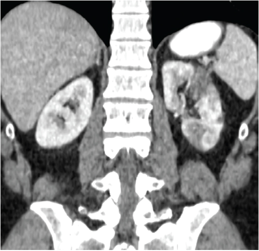
Note multiple hypodensities in the left renal cortex representing renal infarcts.

ad goes here:advert-2
ADVERTISEMENT
SCROLL TO CONTINUE
Common Iliac Artery Dissections
click for large version
Figure 2: Common Iliac Artery Dissections

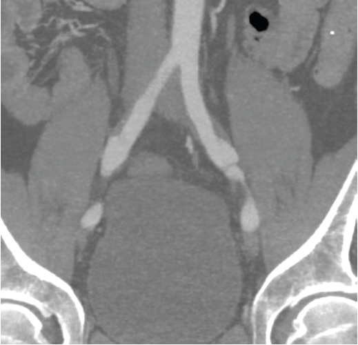
Note bilateral dissections of the common iliac arteries with surrounding soft tissue thickening.

Differential Diagnosis

We were faced with a patient who presented with multifocal dissections with associated inflammatory vessel wall changes as well as elevated inflammatory markers. Treatment with prednisone led to a decrease (although not complete resolution) of inflammatory marker elevations and resolution of some inflammatory changes; however, there was progression of the left common iliac artery dissection. A number of possible etiologies were considered, which can be broadly divided into vasculitides and noninflammatory vasculopathies.

Vasculitis

The most common vasculitis affecting the large vessels in an individual younger than 50 years is Takayasu arteritis (TAK). However, a number of other vasculitides, including Behçet’s and polyarteritis nodosa, can affect the large vessels, such as the renal and iliac arteries. Our patient did not have oral ulcerations to suggest Behçet’s or other signs or symptoms to suggest polyarteritis nodosa. Despite the elevated inflammatory markers, we felt that TAK was less likely because the inflammatory changes on imaging were limited to the areas of dissection and spared other vascular beds affected by TAK, particularly the aorta, subclavian and axillary arteries.1 Although TAK can lead to dissections, the most common arterial lesions are stenoses, which were not seen in our patient. In addition, 80–90% of patients with TAK are women, further decreasing the likelihood of this condition.1

Because our patient’s presentation was not consistent with a primary vasculitis, we felt that the elevated inflammatory markers and inflammatory changes surrounding the vessels on imaging were most likely secondary to the dissections, rather than primary manifestations of the underlying illness. These considerations raised our concern for a noninflammatory vasculopathy as the unifying diagnosis.

Noninflammatory Vasculopathies

Noninflammatory vasculopathies affecting the large vessels are a heterogeneous group of disorders and include Ehler’s Danlos Type IV (EDS IV), Marfan’s syndrome, Loeys-Dietz syndrome, fibromuscular dysplasia (FMD) and segmental arterial mediolysis (SAM). In addition, atherosclerotic lesions can lead to aneurysm and dissection and must be considered in the differential.

Page: 1 2 3 4 5 | Single Page
Share: 

Filed under:ConditionsMeeting ReportsResearch RheumVasculitis Tagged with:AC&RAssociation of Rheumatology Professionals (ARP)AwardsLupus FoundationResearchSalmonTsokos

Related Articles
    Case Report: Ehlers-Danlos Syndrome Mimicking Vasculitis

    Case Report: Ehlers-Danlos Syndrome Mimicking Vasculitis

    July 19, 2018

    A 43-year-old man with a past medical history of type 2 diabetes mellitus, bilateral inguinal hernia repair as a child and prior cholecystectomy woke from sleep with sudden-onset periumbilical abdominal pain. He was admitted to another hospital, but required transfer to our surgical intensive care unit after a recurrent episode of severe abdominal pain, during…

    Fellow’s Forum Case Report: Takayasu’s Arteritis

    May 1, 2014

    How this rare form of large-vessel vasculitis affects different portions, branches of aorta and ways to diagnose, treat and manage the disease

    Study Finds Tocilizumab Could Be Treatment Option for Takayasu Arteritis

    August 17, 2018

    For patients with refractory Takayasu arteritis (TAK), glucocorticoids (GCs) are often provided as the initial therapy for treatment. However, GCs are often associated with adverse effects for long-term use; relapse also occurs frequently during GC tapering.1 TAK involves interleukin (IL) 6. Tocilizumab—a recombinant, humanized, anti-IL-6 receptor (IL-6R) monoclonal antibody—was first reported by Nishimoto et al….

    Lost and found

    The History of ACE Inhibitors in Scleroderma Renal Crisis

    February 16, 2021

    Scleroderma renal crisis is a true medical emergency in rheumatology, one that requires prompt diagnosis and treatment. Here, we review the historic introduction of the angiotensin-converting enzyme inhibitors in this context, and highlight management and key questions moving forward. Background Awareness of renal disease in scleroderma dates back many years. The revered physician William Osler…

  • About Us
  • Meet the Editors
  • Issue Archives
  • Contribute
  • Advertise
  • Contact Us
  • Copyright © 2025 by John Wiley & Sons, Inc. All rights reserved, including rights for text and data mining and training of artificial technologies or similar technologies. ISSN 1931-3268 (print). ISSN 1931-3209 (online).
  • DEI Statement
  • Privacy Policy
  • Terms of Use
  • Cookie Preferences