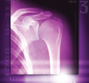
A colored X-ray of a calcified shoulder joint reveals a deposit of calcium at the top right of the shoulder joint.
Miriam Maslo / Science Source
MADRID—Calcification in osteoarthritis (OA) involves a series of pathways and interactions that feed off each other in a process that bears some resemblance to the transformation of cartilage to bone that takes place in the embryonic stage of human development, a researcher said here at the 2017 Annual European Congress on Rheumatology (EULAR).
“My hypothesis is that osteoarthritis is a restart of endochondral ossification in the adult organism,” said Jessica Bertrand, PhD, professor of orthopedic surgery at Otto-von Guericke University in Madgeburg, Germany. “The chondrocytes seem to differentiate into a more hypertrophic state, similar to endochondral bone formation.”
