Video: Every Case Tells a Story| Webinar: ACR/CHEST ILD Guidelines in Practice

An official publication of the ACR and the ARP serving rheumatologists and rheumatology professionals

  • Conditions
    • Axial Spondyloarthritis
    • Gout and Crystalline Arthritis
    • Myositis
    • Osteoarthritis and Bone Disorders
    • Pain Syndromes
    • Pediatric Conditions
    • Psoriatic Arthritis
    • Rheumatoid Arthritis
    • Sjögren’s Disease
    • Systemic Lupus Erythematosus
    • Systemic Sclerosis
    • Vasculitis
    • Other Rheumatic Conditions
  • FocusRheum
    • ANCA-Associated Vasculitis
    • Axial Spondyloarthritis
    • Gout
    • Psoriatic Arthritis
    • Rheumatoid Arthritis
    • Systemic Lupus Erythematosus
  • Guidance
    • Clinical Criteria/Guidelines
    • Ethics
    • Legal Updates
    • Legislation & Advocacy
    • Meeting Reports
      • ACR Convergence
      • Other ACR meetings
      • EULAR/Other
    • Research Rheum
  • Drug Updates
    • Analgesics
    • Biologics/DMARDs
  • Practice Support
    • Billing/Coding
    • EMRs
    • Facility
    • Insurance
    • QA/QI
    • Technology
    • Workforce
  • Opinion
    • Patient Perspective
    • Profiles
    • Rheuminations
      • Video
    • Speak Out Rheum
  • Career
    • ACR ExamRheum
    • Awards
    • Career Development
  • ACR
    • ACR Home
    • ACR Convergence
    • ACR Guidelines
    • Journals
      • ACR Open Rheumatology
      • Arthritis & Rheumatology
      • Arthritis Care & Research
    • From the College
    • Events/CME
    • President’s Perspective
  • Search

Proptosis in Systemic Lupus Erythematosus

Waleed A. Hassan, MD, Samy A. Abdelnaby, MSc, & Yasser M. Abdelhady, MSc  |  Issue: February 2015  |  February 1, 2015

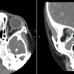
A 30-year-old woman with lupus complained of right eye protrusion and pain.

Presentation

A 30-year-old female patient with systemic lupus erythematosus (SLE) was admitted to the hospital because of a three-day history of protrusion of her right eye and mild right eye pain. There were no other visual complaints. She described polyarthralgia and fatigue. She was six months postpartum, and her pregnancy was without any complication.

She had been diagnosed with lupus three years earlier, and her condition had been well controlled with prednisone and hydroxychloroquine. The patient had stopped taking prednisone following the delivery of her baby.

ad goes here:advert-1
ADVERTISEMENT
SCROLL TO CONTINUE

The physical examination revealed the patient’s temperature was 36.4°C (97.6°F), blood pressure was 130/80 mmHg, pulse rate was

85/min and respiration rate was 18/min. The eye examination showed right eye proptosis, and, apart from slight limitation of upward gaze, there was no limitation of eye movement. The small joints of both hands and her left knee were tender and swollen, and there was trace bilateral ankle edema. There was no skin rash or mouth ulcers. The remainder of the examination was normal.

ad goes here:advert-2
ADVERTISEMENT
SCROLL TO CONTINUE

The erythrocyte sedimentation rate (ESR) was 90 mm/hr. The hemoglobin was 12 g/dL, total white cell count was 3,500/mm3 and the platelet count was 252,000/mm3. Urine analysis disclosed 2+ protein; 1+ blood; 2–3 leukocytes, 3–5 erythro­cytes/high-power field. Urine protein-creatinine ratio was 1.2 mg/dL, albumin was 3.1 g/dL, and the liver enzymes were normal. Serology tests revealed a positive antinuclear antibody (titer of 1:640) and double-stranded DNA 130 IU/mlL (normal <25 IU/mL). The serum anticardiolipin IgM, IgG and lupus anticoagulant were all negative. Serum complement (C3 and C4) levels were decreased. C3 was 65 mg/dL (normal 88–206 mg/dL), and the C4 was 10 mg/dL (normal range 13–75 mg/dL). Blood cultures and thyroid function tests were normal.

A renal biopsy revealed World Health Organization class III lupus nephritis.

A contrast-enhanced computed tomographic (CT) scan of her orbits was consistent with bilateral orbital pseudotumor with more prominent changes on the right (see Figure 1, above). Proptosis due to pseudo-orbital tumor was considered a part of a lupus flare, and pulse steroids in the form of intravenous methylprednisolone (1 gm) were given daily for three days, followed by high-dose oral prednisone at 1 mg/kg/day.

There was dramatic improvement of the eye swelling, arthralgias, leukopenia and proteinuria. The patient has been in remission with a treatment regimen including mycophenolate mofetil (2 gm/day), hydroxychloroquine (400 mg/day) and low-dose prednisone (10 mg/day).

Page: 1 2 3 | Single Page
Share: 

Filed under:ConditionsSystemic Lupus Erythematosus Tagged with:Systemic lupus erythematosus

Related Articles

    Case Report: GPA or IgG4-Related Disease in an 11-Year-Old Girl?

    April 7, 2023

    Granulomatosis with polyangiitis (GPA) is a type of anti-neutrophil cytoplasmic antibody (ANCA) associated small vessel vasculitis that typically affects the kidneys, lungs and sinuses.1 Due to an overlap in signs and symptoms, GPA may initially be difficult to distinguish from IgG4-related disease, another condition that can affect multiple tissues and has variable presentations. Further complicating…

    The Granulomatosis of Wegener’s

    May 16, 2011

    Delving deeper into the nonvasculitis aspects of the disease

    Case Study: 66-Year-Old Man with Visual Loss, Headache, Hematuria, Chronic Sinusitis

    May 8, 2012

    A 66-year-old man presented to the hospital with left-sided visual loss, headache, hematuria, and symptoms of a chronic sinusitis.

    Watch Those Eyes

    December 1, 2007

    What you need to know about Uveitis in Rheumatic Diseases

  • About Us
  • Meet the Editors
  • Issue Archives
  • Contribute
  • Advertise
  • Contact Us
  • Copyright © 2025 by John Wiley & Sons, Inc. All rights reserved, including rights for text and data mining and training of artificial technologies or similar technologies. ISSN 1931-3268 (print). ISSN 1931-3209 (online).
  • DEI Statement
  • Privacy Policy
  • Terms of Use
  • Cookie Preferences