Video: Every Case Tells a Story| Webinar: ACR/CHEST ILD Guidelines in Practice

An official publication of the ACR and the ARP serving rheumatologists and rheumatology professionals

  • Conditions
    • Axial Spondyloarthritis
    • Gout and Crystalline Arthritis
    • Myositis
    • Osteoarthritis and Bone Disorders
    • Pain Syndromes
    • Pediatric Conditions
    • Psoriatic Arthritis
    • Rheumatoid Arthritis
    • Sjögren’s Disease
    • Systemic Lupus Erythematosus
    • Systemic Sclerosis
    • Vasculitis
    • Other Rheumatic Conditions
  • FocusRheum
    • ANCA-Associated Vasculitis
    • Axial Spondyloarthritis
    • Gout
    • Psoriatic Arthritis
    • Rheumatoid Arthritis
    • Systemic Lupus Erythematosus
  • Guidance
    • Clinical Criteria/Guidelines
    • Ethics
    • Legal Updates
    • Legislation & Advocacy
    • Meeting Reports
      • ACR Convergence
      • Other ACR meetings
      • EULAR/Other
    • Research Rheum
  • Drug Updates
    • Analgesics
    • Biologics/DMARDs
  • Practice Support
    • Billing/Coding
    • EMRs
    • Facility
    • Insurance
    • QA/QI
    • Technology
    • Workforce
  • Opinion
    • Patient Perspective
    • Profiles
    • Rheuminations
      • Video
    • Speak Out Rheum
  • Career
    • ACR ExamRheum
    • Awards
    • Career Development
  • ACR
    • ACR Home
    • ACR Convergence
    • ACR Guidelines
    • Journals
      • ACR Open Rheumatology
      • Arthritis & Rheumatology
      • Arthritis Care & Research
    • From the College
    • Events/CME
    • President’s Perspective
  • Search

Race May Not Be a Risk Factor for Giant Cell Arteritis

Lara C. Pullen, PhD  |  October 21, 2019

Although rheumatologists do not know the cause of giant cell arteritis (GCA), researchers have identified many of its risk factors. These factors have largely been determined based on cohorts of patients of Northern European descent that include information about age, sex and HLA DRB1. These past studies have also suggested GCA is uncommon in black patients.

New Findings
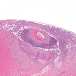
Although biopsy-proven GCA occurs more commonly in women, recent research indicates  its rates are similar between races. The research by Anna M. Gruener, BMBS, an ophthalmologist from Nottingham University Hospitals National Health Service Trust, U.K., and colleagues suggests GCA does not occur more frequently in white patients than in black patients. The investigators published their findings online Aug. 8 in JAMA Ophthalmology. The retrospective cohort study compared the incidence of biopsy proven GCA in black patients with white patients.1

ad goes here:advert-1
ADVERTISEMENT
SCROLL TO CONTINUE

“In our 10-year retrospective study of GCA at the Johns Hopkins Wilmer Eye Institute, we reviewed the medical records of 586 patients who had [temporal artery biopsies], 93 of whom had positive findings,” write the authors in their discussion. “To our knowledge, the only other North American study of GCA incidence with a comparable sample size to ours evaluated patients from Olmsted County, Minn., where the population is predominantly white. That study identified 173 incident cases of GCA during a 50-year study period. Our study is the largest, to our knowledge, to examine GCA incidence in a population that includes a sizeable proportion of black patients.”

The researchers calculated an overall age- and sex-adjusted incidence rate of biopsy-proven GCA of 3.5 (95% confidence interval [CI], 2.5–4.5) per 100,000 population per year.

ad goes here:advert-2
ADVERTISEMENT
SCROLL TO CONTINUE

The Patients
The investigators evaluated patients seen at the Johns Hopkins Wilmer Eye Institute, Baltimore. Patients were selected for temporal artery biopsies based on their clinical presentation. Although the population in the current study differed from previous studies with regards to ethnicity, it was similar to previous studies in terms of mean age (70.5 years) and sex ratios (72.2% women). The mean age of patients selected for temporal artery biopsy was higher in 31 white patients than black patients, mean difference of 2.7 years (95% CI, 0.6–4.7 years; P=0.01).

The researchers classified patients with biopsies of healed arteritis as having biopsy-proven GCA. For this classification, the healing could be a significant fragmentation and/or loss of internal elastic lamina with associated thinning and fibrosis or scarring of the media, sometimes with capillary neovascularization. The researchers classified indeterminate findings of biopsies as negative.

Page: 1 2 3 | Single Page
Share: 

Filed under:ConditionsVasculitis Tagged with:GCAgiant cell arteritis (GCA)raceRisk Factors

Related Articles

    Two Inflammatory Conditions—Polymyalgia Rheumatica and Giant Cell Arteritis—Share Clinical Connection

    March 1, 2013

    Polymyalgia rheumatica (PMR) and giant cell arteritis (GCA) have common clinical and epidemiologic links, but they need not occur synchronously

    Atypical Giant Cell Arteritis Case Illustrates Diagnosis, Management Challenges

    Atypical Giant Cell Arteritis Case Illustrates Diagnosis, Management Challenges

    November 14, 2021

    Giant cell arteritis (GCA) is a granulomatous vasculitis of large- and medium-sized arteries, usually affecting the cranial branches of the aortic arch. It is the most common vasculitis, with the highest risk factor being age. Accurate diagnosis and prompt initiation of therapy are of great importance to prevent serious complications, with the most feared being…

    Updates on Giant Cell Arteritis

    March 19, 2018

    SAN DIEGO—Recent research tells us more about giant cell arteritis (GCA) to help rheumatologists more accurately diagnose and effectively treat patients with this type of vasculitis. On Nov. 6 at the ACR/ARHP Annual Meeting, three experts explored the latest findings on GCA pathogenesis, diagnostic approaches, imaging modalities and growing treatment options. GCA: What’s Really Happening?…

    Experts Discuss Proposed Giant Cell Arteritis Risk Tool

    April 26, 2018

    A proposed model to predict the risk of giant cell arteritis (GCA) prior to a temporal artery biopsy could help triage patients and guide decision making about the need for biopsy or monitoring (see Figure 1). There’s no specific biomarker for GCA, and GCA can be a “diagnostic conundrum, especially when it presents in an…

  • About Us
  • Meet the Editors
  • Issue Archives
  • Contribute
  • Advertise
  • Contact Us
  • Copyright © 2025 by John Wiley & Sons, Inc. All rights reserved, including rights for text and data mining and training of artificial technologies or similar technologies. ISSN 1931-3268 (print). ISSN 1931-3209 (online).
  • DEI Statement
  • Privacy Policy
  • Terms of Use
  • Cookie Preferences