Video: Knock on Wood| Webinar: ACR/CHEST ILD Guidelines in Practice
fa-facebookfa-linkedinfa-youtube-playfa-rss

An official publication of the ACR and the ARP serving rheumatologists and rheumatology professionals

  • Conditions
    • Axial Spondyloarthritis
    • Gout and Crystalline Arthritis
    • Myositis
    • Osteoarthritis and Bone Disorders
    • Pain Syndromes
    • Pediatric Conditions
    • Psoriatic Arthritis
    • Rheumatoid Arthritis
    • Sjögren’s Disease
    • Systemic Lupus Erythematosus
    • Systemic Sclerosis
    • Vasculitis
    • Other Rheumatic Conditions
  • FocusRheum
    • ANCA-Associated Vasculitis
    • Axial Spondyloarthritis
    • Gout
    • Lupus Nephritis
    • Psoriatic Arthritis
    • Rheumatoid Arthritis
    • Systemic Lupus Erythematosus
  • Guidance
    • Clinical Criteria/Guidelines
    • Ethics
    • Legal Updates
    • Legislation & Advocacy
    • Meeting Reports
      • ACR Convergence
      • Other ACR meetings
      • EULAR/Other
    • Research Rheum
  • Drug Updates
    • Analgesics
    • Biologics/DMARDs
  • Practice Support
    • Billing/Coding
    • EMRs
    • Facility
    • Insurance
    • QA/QI
    • Technology
    • Workforce
  • Opinion
    • Patient Perspective
    • Profiles
    • Rheuminations
      • Video
    • Speak Out Rheum
  • Career
    • ACR ExamRheum
    • Awards
    • Career Development
  • ACR
    • ACR Home
    • ACR Convergence
    • ACR Guidelines
    • Journals
      • ACR Open Rheumatology
      • Arthritis & Rheumatology
      • Arthritis Care & Research
    • From the College
    • Events/CME
    • President’s Perspective
  • Search

Reactive Arthritis: Chronic or Self-Limiting?

Michael J. Cammarata, MD  |  Issue: June 2025  |  June 8, 2025

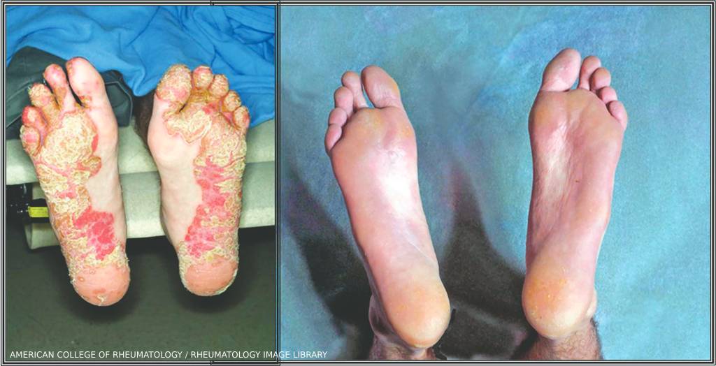
Reactive Arthritis: Keratoderma blennorrhagicum on both feet before (left) and after treatment with a TNFα antagonist. (Click to enlarge.)

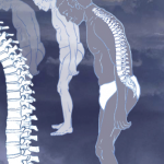
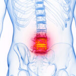
Reactive arthritis is a sterile inflammatory arthritis that often occurs in the weeks following a gastrointestinal or genitourinary tract infection. A form of spondyloarthritis, it can present with myriad extra-articular features and has a variable, sometimes protracted, course that may warrant long-term immunosuppression.

Due to varied presentations, numerous inciting organisms and a lack of specific biomarkers, reactive arthritis is challenging to categorize and study intensively.1 Therefore, management recommendations are often derived from case series and case reports.

ad goes here:advert-1
ADVERTISEMENT
SCROLL TO CONTINUE

In this review, we explore the epidemiology, clinical manifestations, management and prognosis of reactive arthritis, with insights and pearls from experts in the realm of spondyloarthritis.

Historical Context

The combination of arthritis and gastrointestinal or genitourinary symptoms has long been recognized but poorly understood. Arthritis and urethritis were first described together in the 16th century, although identification of culprit organisms, such as chlamydia, didn’t occur until nearly three centuries later.2

ad goes here:advert-2
ADVERTISEMENT
SCROLL TO CONTINUE

In 1916, French physicians Noel Fiessinger and Edgar Leroy described a “syndrome conjunctivo-ureto-synovial” that had occurred in four soldiers after an outbreak of dysentery.3 Four days later, Hans Conrad Julias Reiter described a similar illness, characterized by the triad of arthritis, conjunctivitis and non-gonococcal urethritis. Initially called Reiter’s syndrome, the condition was renamed reactive arthritis given Reiter’s allegiance to the Nazi Party in the 1930s, along with the recognition that a majority of patients do not present with the triad of signs and symptoms he described.4

Epidemiology

The incidence of reactive arthritis varies widely in outbreak studies, but is generally low, around 1% or less, and reported at a global frequency of 0.3–30 per 10,000.5,6 It commonly presents in adults 20–40 years old, with an equal sex distribution; however, reactive arthritis due to Chlamydia trachomatis is more common in men.7

The incidence of HLA-B27 positivity varies widely, with either a small or no increase in frequency reported in outbreak studies and epidemiological surveys, but up to 60–80% reported in hospital-based studies.8,9 HLA-B27 positivity is known to predict a more severe and chronic course, as well as the presence of more extra-articular manifestations. 10 HLA-B27 is, therefore, considered a prognostic tool more than a marker of susceptibility.11

A family history of spondyloarthritis is considered an additional risk factor for chronicity and joint damage.12

The spondyloarthropathies encompass a group of related rheumatic diseases characterized by arthritis, enthesitis and dactylitis with variable degrees of peripheral and axial involvement. These diseases include radiographic and nonradiographic axial spondyloarthritis, psoriatic arthritis, undifferentiated peripheral spondyloarthritis, inflammatory bowel disease-associated arthritis (i.e., enteropathic arthritis) and reactive arthritis (see Figure 1).13

Figure 1. Click to enlarge.

Alexis Ogdie, MD, MSCE, associate professor of medicine and associate professor of epidemiology, Department of Medicine, Hospital of the University of Pennsylvania, Philadelphia, as well as an expert in spondyloarthritis, estimates she has seen only a few cases of reactive arthritis among a larger cohort of patients with spondyloarthritis, which is reflected in the rarity of reactive arthritis represented in previously published spondyloarthritis registries.14

With no validated diagnostic criteria, a diagnosis of reactive arthritis is based on clinical factors. The most widely used criteria for reactive arthritis were proposed by the ACR in 1999 and Braun et al. in 2000.7 The more recent Assessment of SpondyloArthritis International Society (ASAS) classification criteria for spondyloarthritis do not formally include reactive arthritis; however, the classification criteria for peripheral spondyloarthritis include preceding infection.15

Clinical Presentation

Reactive arthritis usually presents as an acute, asymmetric oligoarthritis one to two weeks after onset of a gastrointestinal or genitourinary infection, but can occur up to four weeks after infection with Chlamydia trachomatis.5 Common precipitating organisms include gut bacteria, such as Yersinia, Salmonella, Shigella and Campylobacter, and urogenital tract organisms, such as Chlamydia, Ureaplasma and Mycoplasma. Additional culprit organisms are highlighted in Table 1.5

Click to enlarge.

At disease onset, analysis of synovial fluid can help exclude septic arthritis. Synovial fluid will be inflammatory, with 2,000–64,000 white blood cells, but synovial cultures will be negative.5 Evidence of preceding infection should be sought, although no definite recommended laboratory testing has been established, and often the precipitating infection may go undetected due to lack of symptoms. In fact, a prior infection is identified in only 60% of patients clinically diagnosed with reactive arthritis.16

John Miller, MD, assistant professor of medicine at the Johns Hopkins Arthritis Center, Baltimore, says “the clinical presentation (e.g., diarrhea, hematochezia, dysuria) will often guide the evaluation for infectious etiology. This usually includes stool or urine cultures and nucleic acid testing; however, many other pathogens have been implicated in reactive arthritis (e.g., Giardia, Strongyloides, Clostridium difficile), so additional testing may be necessary, depending on the initial symptoms.”

In keeping with the features of the spondyloarthopathies, reactive arthritis classically presents with an oligoarthritis, favoring the large joints of the lower extremities. However, polyarticular and small joint involvement is also possible. Dactylitis can be seen in up to 40% of patients, and enthesitis is common at the plantar fascia and Achilles tendon.17 Axial involvement is more common in patients who are HLA-B27 positive.11 Importantly, unilateral axial involvement without peripheral arthritis is highly atypical and suggestive of infection.18

Figure 2. Numerous pustules are present on the feet of a patient with reactive arthritis. They begin as vesicles on erythematous bases and become sterile pustules. On the patient’s right sole, the development of keratotic scales (keratoderma blennorrhagica) is most evident. (Click to enlarge.)

Mucocutaneous manifestations are seen in up to 40% of patients.11 Two characteristic manifestations include keratoderma blenorrhagicum and circinate balanitis. The former is characterized by a hyperkeratotic, psoriasiform rash on the palms and soles (see Figure 2), and the latter features shallow, painless serpinginous ulcerations on the glans penis. Dysuria and penile discharge may also be present. Less specific cutaneous signs include aphthous ulcers and erythema nodusum, both of which can also be seen in inflammatory bowel disease (IBD).

Ocular manifestations include a sterile conjunctivitis in up to 50–75% of patients, as well as anterior uveitis/iritis in 12% of patients.11 Much like in axial spondyloarthritis, uveitis can be a chronic, recurring manifestation warranting long-term immunosuppression.

Cardiac manifestations are less common, and seen in up to 10% of patients. Manifestations include aortitis and aortic insufficiency, conduction abnormalities and pericarditis, particularly in patients with a longer duration of illness.11

There are no specific laboratory tests to confirm the diagnosis. Inflammatory markers, such as erythrocyte sedimentation rate (ESR) and C-reactive protein, are typically elevated, with thrombocytosis and anemia of chronic disease reflecting unchecked inflammation. Exclusion of other rheumatic and infectious diseases is paramount. Gonococcal arthritis, rheumatoid arthritis, spondyloarthropathies (e.g., IBD-associated arthritis and psoriatic arthritis), Lyme disease, viral arthritides, Behçet’s disease and crystalline arthropathies should all be investigated, depending on the patient’s age, history and additional clinical features.

Prognosis

Reactive arthritis is often self-limited, with a good prognosis, resolving over an average of three to five months. About one-third of patients recover fully; however, up to half of patients progress to chronic arthritis.19 Overall, the prognosis of an enteric-induced reactive arthritis seems more favorable than one secondary to infection with Chlamydia.11

Dr. Miller

Dr. Miller says reactive arthritis “provides a unique opportunity to explore the complex interplay between infection and autoimmunity, particularly trying to understand why only a fraction of patients develop chronic symptoms.”

Dr. Ogdie notes that due to the self-limited nature of the disease, “by the time patients get to us, it has resolved. However, once diagnosed, patients should be followed intermittently over time to see if they develop a chronic arthritis. I might see a patient with reactive arthritis every year for a couple of years, then increase the interval to every three years, although patients with self-limited disease are often lost to follow-up.

“If the disease is persisting,” Dr. Ogdie stressed, “do a full evaluation for spondyloarthritis and treat it as such. Patients who are HLA-B27 positive with a severe and protracted course are simply patients who have spondyloarthritis that is revealing itself. If I had a magic wand, I would want to know, ‘Is this an isolated episode of arthritis, or will this become a more chronic inflammatory arthritis?’ Time is all we have to sort that out.”

Management

Dr. Ogdie

The primary goal of treatment should start with the prompt identification and treatment of the triggering infection.11 Treatment of Chlamydia is known to reduce the risk of developing reactive arthritis from about 37% to 10%.20 Treatment of sexual partners is also essential to prevent re-infection.

The role for antibiotics after an enteric infection is less clear. At present, antibiotics are not recommended for reactive arthritis secondary to gastrointestinal infection, but can be considered in cases of severe diarrhea, in immunocompromised or elderly patients, or in patients with prior reactive arthritis susceptible to relapse.21

After identification of the antecedent infection, the next step is to provide relief of acute symptoms and minimize the risk of joint and organ damage and disability. Depending on the spectrum of manifestations, disease management may need to be coordinated with a team of specialists, including dermatologists, ophthalmologists and cardiologists.

Non-steroidal anti-inflammatory drugs (NSAIDs) are first-line therapy for musculoskeletal manifestations of reactive arthritis. These can be given at high doses, and agents with a long half-life are preferred for patients with axial involvement.

Intra-articular glucocorticoids can be helpful for cases of mono- or oligo-arthritis, as well as enthesopathy. Oral glucocorticoids are generally reserved for more severe polyarticular involvement or high-risk manifestations, such as severe cardiac disease.

Lucchino et al. recommend a three- to six-month period of observation before consideration of treatment with a conventional synthetic disease-modifying anti-rheumatic drug (DMARD) to avoid treating self-limiting disease.11 Sulfasalazine has been the most studied among these options and has been demonstrated to induce remission more rapidly than placebo. It can be used in chronic disease, but is only effective for peripheral, rather than axial disease.22

If sulfasalazine is contradicted due to G6PD deficiency or sulfonamide allergy, for example, or demonstrates a lack of efficacy, DMARDs, such as azathioprine or methotrexate, can be considered.

“When reactive arthritis becomes chronic,” says Dr. Miller, “its management begins to align closely with other spondyloarthropathies. Use DMARDs and/or biologic immunosuppression, guided by disease severity, axial and extra-articular manifestations.”

Biologic agents have been studied in small case series and case reports. Although there was initial concern regarding the possibility of bacterial proliferation in the setting of tumor necrosis factor (TNF) inhibition, available data suggest biologic efficacy without the risk of precipitating infection.11

Looking Ahead

Reactive arthritis, although rare, remains a significant diagnostic and therapeutic challenge due to its diverse presentation and unpredictable course.

Gaps in knowledge and areas for future study include the lack of diagnostic criteria and treatment guidelines, absence of biomarkers for disease activity and diagnosis, and limited data in long-term clinical trials, particularly related to biologic DMARDs.23 Additional areas of potential study include examining the role of the microbiome in disease pathogenesis and risk factors for progression to chronic spondyloarthritis.24,25

Expanding research efforts can help bridge gaps in knowledge and optimize care for this complex disease.


Michael Cammarata, MD, is an assistant professor of medicine at the Johns Hopkins School of Medicine, Baltimore.

References   

  1. García-Kutzbach A, Chacón-Súchite J, García-Ferrer H, Iraheta I. Reactive arthritis: Update 2018. Clin Rheumatol. 2018 Apr;37(4):869–874.
  2. van Foreest P. (cited by Toivanen A; 2003) Reactive arthritis: Clinical features and treatment. In: Hochberg MC, Silman AJ, Smolen JS, Weinblatt ME, Weisman MH (eds). Rheumatology, Section 9, 3rd ed., vol 2. Spondyloarthropathies. Edinburgh: Mosby, pp. 1233–1240.
  3. Fiessinger N, Leroy E. Contribution à l’etude d’une epidemie de dysenterie dans la Somme. Bull Mem Soc Med Hop Paris. 1916 Juil–Oct;40:2030–2069.
  4. Keitel W. [Hans Reiter and the oculo-urethro-synovial syndrome. 3. The unknown Hans Reiter, scientist and national socialism propagandist.] [Article in German.] Z Rheumatol. 2004 Jun;63(3):244–249.
  5. Hannu T. Reactive arthritis. Best Pract Res Clin Rheumatol. 2011 Jun;25(3):347–357.
  6. Agarwal A, Maikap D, Padhan P. Treatment of reactive arthritis with biological agents. Curr Rheumatol Rep. 2024 Dec;26(12):450–458.
  7. Selmi C, Gershwin ME. Diagnosis and classification of reactive arthritis. Autoimmun Rev. 2014 Apr–May;13(4-5):546–549.
  8. Leirisalo-Repo M, Hannu T, Mattila L. Microbial factors in spondyloarthropathies: Insights from population studies. Curr Opin Rheumatol. 2003 Jul;15(4):408–412.
  9. Aho K, Ahvonen P, Lassus A, et al. HL-A antigen 27 and reactive arthritis. Lancet. 1973 Jul 21;2(7821):157.
  10. Leirisalo M, Skylv G, Kousa M, et al. Followup study on patients with Reiter’s disease and reactive arthritis, with special reference to HLA-B27. Arthritis Rheum. 1982 Mar;25(3):249–259.
  11. Lucchino B, Spinelli FR, Perricone C, et al. Reactive arthritis: Current treatment challenges and future perspectives. Clin Exp Rheumatol. 2019 Nov–Dec;37(6):1065–1076.
  12. Hannu T, Inman R, Granfors K, et al. Reactive arthritis or post-infectious arthritis? Best Pract Res Clin Rheumatol. 2006 Jun;20(3):419–433.
  13. Proft F, Poddubnyy D. Ankylosing spondylitis and axial spondyloarthritis: Recent insights and impact of new classification criteria. Ther Adv Musculoskelet Dis. 2018 Jun;10(5–6):129–139.
  14. Collantes E, Zarco P, Muñoz E, et al. Disease pattern of spondyloarthropathies in Spain: Description of the first national registry (REGISPONSER) extended report. Rheumatology (Oxford). 2007 Aug;46(8):1309–1315.
  15. Rudwaleit M, van der Heijde D, Landewé R, et al. The Assessment of Spondyloarthritis International Society classification criteria for peripheral spondyloarthritis and for spondyloarthritis in general. Ann Rheum Dis. 2011 Jan;70(1):25–31.
  16. Fendler C, Laitko S, Sörensen H, et al. Frequency of triggering bacteria in patients with reactive arthritis and undifferentiated oligoarthritis and the relative importance of the tests used for diagnosis. Ann Rheum Dis. 2001 Apr;60(4):337–343.
  17. Schmitt SK. Reactive Arthritis. Infect Dis Clin North Am. 2017 Jun;31(2):265–277.
  18. Quinzaños-Alonso I. “Reactive Arthritis” in Rheumatology Secrets (West S, Kolfenbach J, eds). Philadelphia: Elsevier, 2019 Dec 8.
  19. Thomson GT, DeRubeis DA, Hodge MA, et al. Post-Salmonella reactive arthritis: Late clinical sequelae in a point source cohort. Am J Med. 1995 Jan;98(1):13–21.
  20. Bardin T, Enel C, Cornelis F, et al. Antibiotic treatment of venereal disease and Reiter’s syndrome in a Greenland population. Arthritis Rheum. 1992 Feb;35(2):190–194.
  21. Hamdulay SS, Glynne SJ, Keat A. When is arthritis reactive? Postgrad Med J. 2006 Jul;82(969):446–453.
  22. Clegg DO, Reda DJ, Weisman MH, et al. Comparison of sulfasalazine and placebo in the treatment of reactive arthritis (Reiter’s syndrome). A Department of Veterans Affairs cooperative study. Arthritis Rheum. 1996 Dec;39(12):2021–2027.
  23. Mullokulov J. Reactive arthritis [unpublished manuscript]. 2024.
  24. Breban M, Tap J, Leboime A, et al. Faecal microbiota study reveals specific dysbiosis in spondyloarthritis. Ann Rheum Dis. 2017 Sep;76(9):1614–1622.
  25. Manasson J, Shen N, Garcia Ferrer HR, et al. Gut microbiota perturbations in reactive arthritis and postinfectious spondyloarthritis. Arthritis Rheumatol. 2018 Feb;70(2):242–254.

Acknowledgment

The author thanks Javohir Mullokulov MD, assistant professor, and Khalmurad Akhmedov, DSc, professor, both of Tashkent Medical Academy, Uzbekistan, for the idea that inspired this article and for their assistance in its development.

Share: 

Filed under:Axial SpondyloarthritisConditions Tagged with:biologic DMARDsdisease managementextra-articular manifestationsGlucocorticoidsHLA-B27infectious etiologymusculoskeletal manifestationsNSAIDsReactive arthritisspondyloarthritissulfasalazine

Related Articles

    Top Research in Axial Spondyloarthritis Presented at ACR Convergence 2022

    November 18, 2022

    PHILADELPHIA—Approximately 100 research abstracts on axial spondyloarthritis (axSpA) were accepted for presentation at ACR Convergence 2022. It is exciting to see a wealth of research on axSpA being undertaken worldwide. Here, we highlight important points from 10 of these studies. 1. Abstract 0378: Prevalence of Axial Spondyloarthritis (axSpA) in Patients Treated for Chronic Back Pain…

    Rheumatologists Make Progress Defining Spectrum of Axial Spondyloarthritis

    May 1, 2014

    New research clarifies terminology used for diagnosis, but questions remain around epidemiology, genetics and management of patients with axial skeletal inflammation

    Nelosa/shutterstock.com

    Men, Women & Medical Differences in Axial Spondyloarthropathy

    October 24, 2019

    Historically, ankylosing spondylitis was considered mainly a male disease. But it has become evident this predominance is not as great as previously believed. Here we discuss recent developments in the area, including potential differences between the sexes in symptom and disease burden, immunological and genetic background, diagnostic delay, treatment response and ongoing research questions. Medical…

    Key Research in Axial Spondyloarthritis Encapsulated

    December 2, 2024

    Our understanding of (axSpA) has really changed, especially with regard to male-female differences. In this report, we identify important research on axSpA presented at ACR Convergence 2024, summarize the abstracts and comment on why each is important, addressing the relevance for clinicians and the potential impact on future research.

  • About Us
  • Meet the Editors
  • Issue Archives
  • Contribute
  • Advertise
  • Contact Us
fa-facebookfa-linkedinfa-youtube-playfa-rss
  • Copyright © 2025 by John Wiley & Sons, Inc. All rights reserved, including rights for text and data mining and training of artificial technologies or similar technologies. ISSN 1931-3268 (print). ISSN 1931-3209 (online).
  • DEI Statement
  • Privacy Policy
  • Terms of Use
  • Cookie Preferences