Video: Knock on Wood| Webinar: ACR/CHEST ILD Guidelines in Practice
fa-facebookfa-linkedinfa-youtube-playfa-rss

An official publication of the ACR and the ARP serving rheumatologists and rheumatology professionals

  • Conditions
    • Axial Spondyloarthritis
    • Gout and Crystalline Arthritis
    • Myositis
    • Osteoarthritis and Bone Disorders
    • Pain Syndromes
    • Pediatric Conditions
    • Psoriatic Arthritis
    • Rheumatoid Arthritis
    • Sjögren’s Disease
    • Systemic Lupus Erythematosus
    • Systemic Sclerosis
    • Vasculitis
    • Other Rheumatic Conditions
  • FocusRheum
    • ANCA-Associated Vasculitis
    • Axial Spondyloarthritis
    • Gout
    • Lupus Nephritis
    • Psoriatic Arthritis
    • Rheumatoid Arthritis
    • Systemic Lupus Erythematosus
  • Guidance
    • Clinical Criteria/Guidelines
    • Ethics
    • Legal Updates
    • Legislation & Advocacy
    • Meeting Reports
      • ACR Convergence
      • Other ACR meetings
      • EULAR/Other
    • Research Rheum
  • Drug Updates
    • Analgesics
    • Biologics/DMARDs
  • Practice Support
    • Billing/Coding
    • EMRs
    • Facility
    • Insurance
    • QA/QI
    • Technology
    • Workforce
  • Opinion
    • Patient Perspective
    • Profiles
    • Rheuminations
      • Video
    • Speak Out Rheum
  • Career
    • ACR ExamRheum
    • Awards
    • Career Development
  • ACR
    • ACR Home
    • ACR Convergence
    • ACR Guidelines
    • Journals
      • ACR Open Rheumatology
      • Arthritis & Rheumatology
      • Arthritis Care & Research
    • From the College
    • Events/CME
    • President’s Perspective
  • Search

Recurrent Medial Elbow Pain Following Successful Tommy John Surgery

Mark H. Greenberg, MD, RMSK, RhMSUS, A. Lee Day, MD, RMSK, James W. Fant Jr., MD, & Christopher G. Mazoue, MD  |  Issue: August 2020  |  August 12, 2020

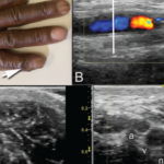
Ulnar Nerve Relocation: A New Syndrome?

Elements of this proposed syndrome would be: sonographic demonstration of ulnar nerve dislocation pushed by a hypertrophied and dislocating MHTr muscle, with pain occurring at the end of rapid elbow extension.

The typical clinical setting would be in a professional athlete who repetitively throws.


References

  1. Barco R, Antuña SA. Medial elbow pain. EFORT Open Rev. 2017Aug 30;2(8):362–371.
  2. Cutts S. Cubital tunnel syndrome. Postgrad Med J. 2007 Jan;83(975):28–31.
  3. Bjerre JJ, Johannsen FE, Rathcke M, Krogsgaard MR. Snapping elbow—A guide to diagnosis and treatment. World J Orthop. 2018 Apr 18;9(4):65–71.
  4. Jacobson JA, Jebson PJ, Jeffers AW, et al. Ulnar nerve dislocation and snapping triceps syndrome: Diagnosis with dynamic sonography—report of three cases. Radiology. 2001 Sep;220(3):601–605.
  5. Jobe FW, Stark H, Lombardo SJ. Reconstruction of the ulnar collateral ligament in athletes. J Bone Joint Surg Am. 1986 Oct;68(8):1158–1163.
  6. Somerson JS, Petersen JP, Neradilek MB, et al. Complications and outcomes after medial ulnar collateral ligament reconstruction: A meta-regression and systematic review. JBJS Rev. 2018 May;6(5):e4.
  7. Leland DP, Conte S, Flynn N, et al. Prevalence of medial ulnar collateral ligament surgery in 6135 current professional baseball players: A 2018 update. Orthop J Sports Med. 2019 Sep 25;7(9):2325967119871442.
  8. Gutierrez NM, Granville C, Kaplan L, et al. Elbow MRI findings do not correlate with future placement on the disabled list in asymptomatic professional baseball pitchers. Sports Health. 2017 May/Jun;9(3):222–229.
  9. Garcia GH, Gowd AK, Cabarcas BC, et al. Magnetic resonance imaging findings of the asymptomatic elbow predict injuries and surgery in Major League Baseball pitchers. Orthop J Sports Med. 2019 Jan 20;7(1):2325967118818413.
  10. Cobb F. IV. Report of a case of recurrent dislocation of the ulnar nerve cured by operation: With summary of previously reported cases. Ann Surg. 1903;38(5):652–663.
  11. Lazaro L 3rd. Ulnar nerve instability: Ulnar nerve injury due to elbow flexion. South Med J. 1977 Jan;70(1):36–40.
  12. Xarchas KC, Psillakis I, Koukou O, et al. Ulnar nerve dislocation at the elbow: Review of the literature and report of three cases. Open Orthop J. 2007 Sep 24;1:1–3.
  13. Spinner RJ, Goldner RD. Snapping of the medial head of the triceps and recurrent dislocation of the ulnar nerve. Anatomical and dynamic factors. J Bone Joint Surg Am. 1998 Feb;80(2):239–247.
  14. Rioux-Forker D, Bridgeman J, Brogan DM. Snapping triceps syndrome. J Hand Surg Am. 2018 Jan;43(1):90.e1–90.e5.
  15. Watts AC, McEachan J, Reid J, Rymaszewski L. The snapping elbow: A diagnostic pitfall. J Shoulder Elbow Surg. 2009 Jan–Feb;18(1):e9–e10.
  16. Spinner RJ, Hayden FR Jr., Hipps CT, Goldner RD. Imaging the snapping triceps. AJR Am J Roentgenol. 1996 Dec;167(6):1550–1551.
ad goes here:advert-1
ADVERTISEMENT
SCROLL TO CONTINUE

Page: 1 2 3 4 5 | Single Page
Share: 

Filed under:Conditions Tagged with:case reportdiagnostic imagingelbow painTommy John Surgeryulnar nerveulnar nerve instabilityulnar nerve relocation syndromeultrasonographyUltrasound

Related Articles

    Ortho Angle

    August 1, 2009

    Where rheumatology and orthopedics meet

    Using Ultrasound to Diagnose Carpal Tunnel Syndrome

    April 26, 2018

    Note: Updated May 2, 2018, to correct a link in the reference section. The error was introduced in editing. A 44-year-old Caucasian woman presented to the outpatient rheumatology clinic that had followed her for several years for rheumatoid arthritis. She was compliant with her regimen of hydroxychloroquine, etanercept and salsalate. Her chief complaint was worsening…

    Clockwise from top: Image A shows the periungual ulceration (arrow) of the fifth digit, as well as the prior amputation of the second digit. Image B shows a longitudinal ultrasound of the palmar aspect of the ulnar right wrist. In the center of the image, the ulnar artery is in view and color Doppler flow is visualized within the vessel until there is a reversal of flow demonstrated by color change, followed by severe attenuation of flow distally. Distal to the cessation of flow, the vessel appears hypoechoic due to proliferation of the intima to the point of occluding the lumen. Image C shows a transverse view at the level of white line in Image B, with endothelial proliferation seen around a central lumen of the artery (a), next to the vein (v) and ulnar nerve (n). Image D, shows a transverse view at the level of the black line in Image B, where the arterial lumen (a) is occluded.

    Insights Into Ulnar Artery Occlusion in Systemic Sclerosis

    April 17, 2021

    A 51-year-old man with a history of limited systemic sclerosis with Raynaud’s phenomenon and pulmonary hypertension being treated with tadalafil and macitentan presented to a clinic with ulceration of his right pinkie. The patient had injured the finger two months earlier. He reported poor healing and the presence of a persistent ulcer since the injury….

    Insights on the Diagnosis & Treatment of Low Back & Hip Pain

    March 19, 2019

    CHICAGO—Two experts presented insights on the diagnosis and treatment of low back and hip pain, including a refresher course on the mechanical structures involved, in Anatomy in a Day: Demystifying Low Back Pain and Lateral Hip Pain: New Patho-Anatomical Perspectives, a session at the 2018 ACR/ARHP Annual Meeting. Low Back Pain Avoid using such terms…

  • About Us
  • Meet the Editors
  • Issue Archives
  • Contribute
  • Advertise
  • Contact Us
fa-facebookfa-linkedinfa-youtube-playfa-rss
  • Copyright © 2025 by John Wiley & Sons, Inc. All rights reserved, including rights for text and data mining and training of artificial technologies or similar technologies. ISSN 1931-3268 (print). ISSN 1931-3209 (online).
  • DEI Statement
  • Privacy Policy
  • Terms of Use
  • Cookie Preferences