Video: Every Case Tells a Story| Webinar: ACR/CHEST ILD Guidelines in Practice

An official publication of the ACR and the ARP serving rheumatologists and rheumatology professionals

  • Conditions
    • Axial Spondyloarthritis
    • Gout and Crystalline Arthritis
    • Myositis
    • Osteoarthritis and Bone Disorders
    • Pain Syndromes
    • Pediatric Conditions
    • Psoriatic Arthritis
    • Rheumatoid Arthritis
    • Sjögren’s Disease
    • Systemic Lupus Erythematosus
    • Systemic Sclerosis
    • Vasculitis
    • Other Rheumatic Conditions
  • FocusRheum
    • ANCA-Associated Vasculitis
    • Axial Spondyloarthritis
    • Gout
    • Psoriatic Arthritis
    • Rheumatoid Arthritis
    • Systemic Lupus Erythematosus
  • Guidance
    • Clinical Criteria/Guidelines
    • Ethics
    • Legal Updates
    • Legislation & Advocacy
    • Meeting Reports
      • ACR Convergence
      • Other ACR meetings
      • EULAR/Other
    • Research Rheum
  • Drug Updates
    • Analgesics
    • Biologics/DMARDs
  • Practice Support
    • Billing/Coding
    • EMRs
    • Facility
    • Insurance
    • QA/QI
    • Technology
    • Workforce
  • Opinion
    • Patient Perspective
    • Profiles
    • Rheuminations
      • Video
    • Speak Out Rheum
  • Career
    • ACR ExamRheum
    • Awards
    • Career Development
  • ACR
    • ACR Home
    • ACR Convergence
    • ACR Guidelines
    • Journals
      • ACR Open Rheumatology
      • Arthritis & Rheumatology
      • Arthritis Care & Research
    • From the College
    • Events/CME
    • President’s Perspective
  • Search

Retroperitoneal Fibrosis & IgG4-Related Disease

Mithu Maheswaranathan, MD  |  December 11, 2024

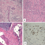
One difference observed in isolated RPF is that the tissue does not have as many IgG4+ plasma cells, but also fewer cells in general. “This can result in lower overall IgG4+ plasma cells, but a higher IgG4/IgG ratio, where almost all plasma cells in a biopsy are IgG4 positive, for example,” Dr. Stone explained.

Also, a subset of non-IgG4-related RPF that is poorly understood to date is characterized on biopsy with acellular fibrosis and small lymphoid aggregates, and sometimes entrapped, necrotic fat cells.

ad goes here:advert-1
ADVERTISEMENT
SCROLL TO CONTINUE

Dr. Stone explained how RPF should be considered on a spectrum with IgG4-RD, with significant overlap in some patients. He provided the example of the polymyalgia rheumatica/giant cell arteritis spectrum as a similar parallel to the association of RPF and IgG4-RD.

Two subtypes of IgG4-RD are a fibrotic subtype and proliferative subtype. The fibrotic subtype has low frequency of atopy, typically normal serologic activity (normal IgG4, eosinophils and complement levels), and pathology with sparse IgG4+ cells per HPF but an IgG4/IgG ratio usually greater than 40%. The proliferative subtype of IgG4-RD has high prevalence of atopy, often serologic activity (elevated IgG4 levels, eosinophilia, hypocomplementemia), high numbers of IgG4+ plasma cells on biopsy in the lymphoplasmacytic infiltrate and very robust response to treatment.

ad goes here:advert-2
ADVERTISEMENT
SCROLL TO CONTINUE

Although RPF is classically grouped under the fibrotic subtype, Dr. Stone explained how some patients will have features similar to IgG4-RD and multisystem organ involvement.

Systemic Involvement Outside the Retroperitoneum

Dr. Stone noted that the traditional approach to RPF, which even he adopted for many years, as “typically confined to the retroperitoneum is really not true.”

In the cohort at Massachusetts General Hospital, Dr. Stone noted they have treated 571 IgG4-RD patients, of which 22% (124) have RPF. Among these cases of RPF, 56 have RPF only and 68 have RPF with other organ involvement. For example, 47% of patients with RPF in the MGH cohort (who fulfill ACR/EULAR classification criteria) have pancreatic disease, 43% have submandibular gland disease, 39% have renal involvement and 25% have aorta or large vessel disease (see Table 2).

“If you look down the list of organs  involved [in RPF], it reads like any large cohort of patients with IgG4-related disease,” Dr. Stone said.

Table 2: Organ Involvement in RPF
Pancreas (47%)
Submandibular Gland (43%)
Kidney (39%)
Aorta/Large Vessels (25%)
Lacrimal Gland (22%)
Lymph Node (22%)
Lungs (20%)

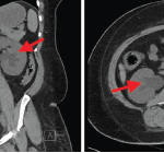
Caution of RPF Mimics, Especially Malignancy

For the latter part of the talk, Dr. Stone reviewed clinical cases that he has managed and offered some cautious pointers for clinicians in managing these patients.

Page: 1 2 3 | Single Page
Share: 

Filed under:ACR ConvergenceConditionsOther Rheumatic Conditions Tagged with:ACR Convergence 2024IgG4 related diseaseIgG4-RDretroperitoneal fibrosis

Related Articles
    IgG4-Related Kidney Disease: Diagnostics, Manifestations, & More

    IgG4-Related Kidney Disease: Diagnostics, Manifestations & More

    May 17, 2018

    Immunoglobin G4-related disease (IgG4-RD) is a rare fibro-inflammatory disease of unknown etiology that has been recently recognized. It can cause fibro-inflammatory masses in almost every organ of the body and is associated with dense lymphoplasmacytic infiltration of IgG4-postitive plasma cells, storiform fibrosis and elevated levels of serum IgG4.1 IgG4-RD is a systemic disease that may…

    A Spotlight on IgG4-Related Disease

    January 1, 2013

    What rheumatologists need to know about identifying and diagnosing immunoglobulin G4-related disease (IgG4-RD)

    Case Report: Atypical Presentation of Idiopathic Retroperitoneal Fibrosis

    March 14, 2022

    Retroperitoneal fibrosis (RPF) is a rare condition characterized by aberrant fibroinflammatory tissue developing in the retroperitoneum. This disorder was initially called Ormond’s disease. RPF may be idiopathic or secondary to other conditions. Idiopathic RPF is a part of the disease spectrum of chronic periaortitis due to its typical periaortoiliac localization. Idiopathic retroperitoneal fibrosis is a…

    Case Report: Sarcoidosis in Patient with History of IgG4-Related Disease

    September 14, 2021

    Sarcoidosis and IgG4-related disease (IgG4-RD) are both immune-mediated, often multi-organ, diseases of uncertain etiology capable of presenting with diverse clinical manifestations. Many clinical features are common to both conditions, including hypergammaglobulinemia, the ability to form inflammatory masses and involvement of the lymph nodes, lacrimal glands, salivary glands, meninges and lungs. Although imaging modalities, such as…

  • About Us
  • Meet the Editors
  • Issue Archives
  • Contribute
  • Advertise
  • Contact Us
  • Copyright © 2025 by John Wiley & Sons, Inc. All rights reserved, including rights for text and data mining and training of artificial technologies or similar technologies. ISSN 1931-3268 (print). ISSN 1931-3209 (online).
  • DEI Statement
  • Privacy Policy
  • Terms of Use
  • Cookie Preferences