Dr. Cutolo
Ophthalmoscopes and traditional microscopes are two, low-cost options that are widely available and can be used with minimal training. Dermatoscopes are portable devices at intermediate cost that allow nailfold capillary viewing. Stereo microscopes are the main tools used in national centers due to their ease of use and intermediate cost. But Stereo microscopes can be difficult to use on patients with joint contractures. Dermatoscopes, ophthalmoscopes and traditional microscopes can provide only low magnification, whereas stereo microscopes and video capillaroscopes can provide either low- or high-level magnification.3,5
A video capillaroscope is the most expensive option. It consists of an optical microscope and a digital video camera combined and connected to a computer. The computer can be used to calculate such measurements as the capillary loop diameter and the intercapillary distance.4 It has a handheld probe that can be used for bedside examination or in patients with severe flexion contractures.2 It is available as a portable system, allowing for easy clinical use. Videocapillaroscopy is considered the gold standard for noninvasive examination of the microcirculation. Currently, relatively few rheumatologists have access to this tool, except at large clinical centers, although this may be changing.3
Assessment of Raynaud’s Syndrome
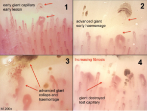
Classical morphological changes of the microvessels (irreversible and pathological) that identify different steps of the sclerodermic microangiopathy (magnification 200X).
Currently, capillaroscopy’s main application is in the assessment of patients with Raynaud’s phenomenon. Raynaud’s phenomenon is characterized by paroxysmal reversible episodes of vasospasm in the peripheral small vessels of the fingers or toes. In primary Raynaud’s phenomenon (about 80% of patients), this occurs without the presence of underlying disease. However, secondary Raynaud’s phenomenon is associated with other medical conditions, most commonly connective tissue disease. In secondary Raynaud’s phenomenon, unlike primary Raynaud’s phenomenon, patients have structural vascular damage that can be viewed via nailfold-viewing methods.7