Video: Every Case Tells a Story| Webinar: ACR/CHEST ILD Guidelines in Practice

An official publication of the ACR and the ARP serving rheumatologists and rheumatology professionals

  • Conditions
    • Axial Spondyloarthritis
    • Gout and Crystalline Arthritis
    • Myositis
    • Osteoarthritis and Bone Disorders
    • Pain Syndromes
    • Pediatric Conditions
    • Psoriatic Arthritis
    • Rheumatoid Arthritis
    • Sjögren’s Disease
    • Systemic Lupus Erythematosus
    • Systemic Sclerosis
    • Vasculitis
    • Other Rheumatic Conditions
  • FocusRheum
    • ANCA-Associated Vasculitis
    • Axial Spondyloarthritis
    • Gout
    • Psoriatic Arthritis
    • Rheumatoid Arthritis
    • Systemic Lupus Erythematosus
  • Guidance
    • Clinical Criteria/Guidelines
    • Ethics
    • Legal Updates
    • Legislation & Advocacy
    • Meeting Reports
      • ACR Convergence
      • Other ACR meetings
      • EULAR/Other
    • Research Rheum
  • Drug Updates
    • Analgesics
    • Biologics/DMARDs
  • Practice Support
    • Billing/Coding
    • EMRs
    • Facility
    • Insurance
    • QA/QI
    • Technology
    • Workforce
  • Opinion
    • Patient Perspective
    • Profiles
    • Rheuminations
      • Video
    • Speak Out Rheum
  • Career
    • ACR ExamRheum
    • Awards
    • Career Development
  • ACR
    • ACR Home
    • ACR Convergence
    • ACR Guidelines
    • Journals
      • ACR Open Rheumatology
      • Arthritis & Rheumatology
      • Arthritis Care & Research
    • From the College
    • Events/CME
    • President’s Perspective
  • Search

Sonographic Diagnosis of Knuckle Pads

Pankaj Bansal, MD, RhMSUS, Eugene Kissin, MD, RhMSUS, & Fawad Aslam, MBBS, RhMSUS, RMSK  |  Issue: October 2022  |  October 10, 2022

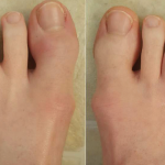
Musculoskeletal ultrasound can be an important tool in confirming the diagnosis of knuckle pads. On gray-mode sonography, knuckle pads are hypoechoic, subcutaneous masses with ill-defined margins that are usually non-compressible.5 Doppler usually does not show any hypervascularity in the nodule, although peripheral hypervascularity has been rarely reported.6 The underlying joint and tendon are usually normal. Histopathology of knuckle pads reveals myofibroblast proliferation and decreased elastic filaments in the dermis. The epidermis and corneum are normal.5

Similar to our case, knuckle pads in association with Dupuytren’s contractures have been reported.2 Dupuytren’s contractures are secondary to fibrous thickening of the palmar fascia, leading to firm palmar nodules and cords. On musculoskeletal ultrasound, these subcutaneous nodules in the palmar fascia are directly superficial to the flexor tendons and appear hypoechoic. Hypervascularity on color Doppler is usually absent and the lesions are non-compressible. Typically, the length of these lesions is greater than the width.7

ad goes here:advert-1
ADVERTISEMENT
SCROLL TO CONTINUE

In contrast, flexor tenosynovitis of the flexor tendons of the hands appears as a hypoechoic or anechoic compressible swelling around the tendon (i.e., visible both above and beyond the tendon) with increased color Doppler signal and is accompanied by thickening and loss of the normal fibrillar pattern of the tendon.8

Gouty tophi can also appear on the dorsal, as well as the volar, aspect of the PIP joints. They are usually soft to firm and non-tender. Sonographically, they are heterogeneous and can be multilobulated.9 They can contain hyperechoic calcifications with acoustic shadowing.10 The underlying joint may reveal a double contour sign secondary to crystalline deposition on the cartilage surface.11 Juxta-articular erosions may also be seen underneath the tophaceous deposits.9,12 Hypervascularity can be seen around the periphery of the tophus.

ad goes here:advert-2
ADVERTISEMENT
SCROLL TO CONTINUE

Rheumatoid nodules are also painless, soft to firm, non-tender nodules and can appear on the dorsal or volar aspect of the PIP joints. Sonographically, they are homogenous hypoechoic nodules with hyperechoic rims and may have anechoic centers.10 Features of synovitis, such as synovial hypertrophy, effusion and hypervascularity with increased color Doppler uptake, may be present. In addition, periarticular erosions can also be seen, although the nodules themselves are less erosive to the bone, and cortical bone is easily seen underneath the nodule.10

Synovitis of the PIP joints can also mimic knuckle pads. In contrast to knuckle pads, synovitis is associated with pain, tenderness and limited mobility of the PIP joint. Ultrasound reveals effusion, synovial hypertrophy and increased color Doppler signal in the PIP joint.

Page: 1 2 3 4 | Single Page
Share: 

Filed under:Conditions Tagged with:case reportDupuytren's contractureknuckle padsUltrasound

Related Articles
    Musculoskeletal Ultrasound: A Valuable Tool for Diagnosing Rheumatic Illnesses

    Musculoskeletal Ultrasound: A Valuable Tool for Diagnosing Rheumatic Illnesses

    October 15, 2015

    Musculoskeletal (MSK) ultrasound is a valuable imaging modality for the practicing rheumatologist and provides an efficient tool with high diagnostic value in the evaluation of patients with musculoskeletal complaints. The use of MSK ultrasound has evolved in the U.S. due to the emergence of less-expensive, portable ultrasound units, which provide high-quality gray-scale and power Doppler…

    Using Ultrasound to Diagnose Carpal Tunnel Syndrome

    April 26, 2018

    Note: Updated May 2, 2018, to correct a link in the reference section. The error was introduced in editing. A 44-year-old Caucasian woman presented to the outpatient rheumatology clinic that had followed her for several years for rheumatoid arthritis. She was compliant with her regimen of hydroxychloroquine, etanercept and salsalate. Her chief complaint was worsening…

    Case Report: A Psoriatic Arthritis Patient with Dactylitis & Enthesitis

    September 20, 2018

    A 36-year-old woman presented at the Johns Hopkins Arthritis Center for a second opinion regarding a diagnosis of psoriatic arthritis (PsA). One year prior to our evaluation, she had developed pain and stiffness in her hands, feet, knees, ankles, elbows and shoulders. She had mild plaque psoriasis of the scalp and base of the neck,…

    Case Report: Perplexing Pulmonary Nodules

    March 14, 2022

    Pulmonary nodules are com­mon; most are benign, but the differential diagnosis is broad and includes life-threatening possibilities.1 Our patient is a former smoker who has a history of a complex autoimmune disease and multiple pulmonary nodules. This case was challenging, but clinical, radiographic and histologic clues helped lead to the correct diagnosis. Case Presentation The…

  • About Us
  • Meet the Editors
  • Issue Archives
  • Contribute
  • Advertise
  • Contact Us
  • Copyright © 2025 by John Wiley & Sons, Inc. All rights reserved, including rights for text and data mining and training of artificial technologies or similar technologies. ISSN 1931-3268 (print). ISSN 1931-3209 (online).
  • DEI Statement
  • Privacy Policy
  • Terms of Use
  • Cookie Preferences