Video: Every Case Tells a Story| Webinar: ACR/CHEST ILD Guidelines in Practice

An official publication of the ACR and the ARP serving rheumatologists and rheumatology professionals

  • Conditions
    • Axial Spondyloarthritis
    • Gout and Crystalline Arthritis
    • Myositis
    • Osteoarthritis and Bone Disorders
    • Pain Syndromes
    • Pediatric Conditions
    • Psoriatic Arthritis
    • Rheumatoid Arthritis
    • Sjögren’s Disease
    • Systemic Lupus Erythematosus
    • Systemic Sclerosis
    • Vasculitis
    • Other Rheumatic Conditions
  • FocusRheum
    • ANCA-Associated Vasculitis
    • Axial Spondyloarthritis
    • Gout
    • Psoriatic Arthritis
    • Rheumatoid Arthritis
    • Systemic Lupus Erythematosus
  • Guidance
    • Clinical Criteria/Guidelines
    • Ethics
    • Legal Updates
    • Legislation & Advocacy
    • Meeting Reports
      • ACR Convergence
      • Other ACR meetings
      • EULAR/Other
    • Research Rheum
  • Drug Updates
    • Analgesics
    • Biologics/DMARDs
  • Practice Support
    • Billing/Coding
    • EMRs
    • Facility
    • Insurance
    • QA/QI
    • Technology
    • Workforce
  • Opinion
    • Patient Perspective
    • Profiles
    • Rheuminations
      • Video
    • Speak Out Rheum
  • Career
    • ACR ExamRheum
    • Awards
    • Career Development
  • ACR
    • ACR Home
    • ACR Convergence
    • ACR Guidelines
    • Journals
      • ACR Open Rheumatology
      • Arthritis & Rheumatology
      • Arthritis Care & Research
    • From the College
    • Events/CME
    • President’s Perspective
  • Search

The Art & Science of Imaging the Pediatric Sacroiliac Joint

Lara C. Pullen, PhD  |  November 3, 2021

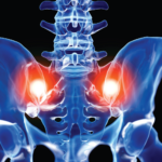
Radiologists and rheumatologists who interpret pediatric pelvic magnetic resonance imaging (MRI) scans must be able to differentiate inflammatory changes from the normal physiologic changes of a maturing sacroiliac (SI) joint. However, this ability varies by specialist, a point highlighted by a new study from Pamela F. Weiss, MD, associate professor of pediatrics and epidemiology, University of Pennsylvania Perelman School of Medicine, Philadelphia, and colleagues. Their findings suggest additional training may be needed for radiologists regarding the appearance of the maturing SI joint on MRI.

Dr. Weiss and colleagues evaluated the agreement between local radiologists’ interpretations of pediatric SI joint MRIs and a central imaging team’s interpretation. Their study was published in the June issue of Arthritis Care & Research.1

ad goes here:advert-1
ADVERTISEMENT
SCROLL TO CONTINUE

Study Details

The study included the evaluation of the SI joints of 120 children performed in eight pediatric hospitals across North America. With a median age of 14 years, the children had a median disease duration of 0.8 years at the time of imaging. Half the children were male, and the majority were white (76.8%). In addition, the majority of children had a diagnosis of enthesitis-related arthritis (82.4%). The investigators coded the global impression of sacroiliitis based on the impression section of the radiology report. If the impression did not differentiate between active inflammation and chronic sacroiliitis, the researchers looked at the findings section of the radiology report. If bone marrow edema in or along the SI joints was noted in the findings section, then the report was coded as chronic inflammation.

The MRIs were independently reviewed by three experienced musculoskeletal pediatric radiologists serving as the central review team for the study. These three radiologists were selected for their ability to identify inflammatory and structural lesions at the SI joint. The majority of the time (84%), the central radiologists agreed on the presence or absence of active inflammation and chronic lesions. The results of the central review team were compared with reports from the children’s local radiologists. The authors note that no standardized assessment for local interpretation exists, and the imaging quality of the studies reviewed was highly variable—even within the same institution.

ad goes here:advert-2
ADVERTISEMENT
SCROLL TO CONTINUE

The local reports had a low, positive predictive value of 51.8%. In other words, only 51.8% of the diagnoses of sacroiliitis made locally were confirmed by the centralized readers. Only rarely (2% of the time) did the central radiologists identify active inflammation when the local radiologists did not identify active inflammation. It was much more common (22.5% of the time) for the local radiologists to identify active inflammation and the central radiologists conclude that there was no active inflammation. Thirteen of the studies were missing coronal oblique sequences, making visualization of the synovial section of the joint more difficult than if the sequences were available. However, when investigators performed a sensitivity analysis that excluded all studies with any quality issues or missing coronal oblique sequences, the rates of discordance were approximately the same.

Page: 1 2 3 | Single Page
Share: 

Filed under:ConditionsPediatric Conditions Tagged with:imagingjoint imagingPediatricRadiologistsacroiliac jointssacroiliitisSI joint

Related Articles

    Imaging of Axial Psoriatic Arthritis

    May 9, 2022

    The axial phenotype of psoriatic arthritis (axPsA) is an excellent example of a major controversy in rheumatology that has become the focus of attention because of the emergence of new therapies with different mechanisms of action for alleviating joint inflammation. It was first described in 1961 but, until recently, it has largely remained under the…

    Imaging in Ankylosing Spondylitis

    April 1, 2015

    MRI inflammation, fat and new bone formation in the sacroiliac joints, spine in patients with AS

    Envision Arthritis Pathology

    April 1, 2008

    MRI advances in RA and OA

    Kateryna Kon / Shutterstock.com

    How to Manage, Treat Anemia of Inflammation in Patients with Rheumatic Disease

    December 17, 2017

    Anemia is common in patients with systemic rheumatic disease, yet it may not get the attention it deserves. Anemia can result from chronic inflammation, treatment side effects or other disease factors, or it may signal an unrelated condition. Although diagnosis and treatment of anemia are sometimes challenging, clinicians must do their utmost to rigorously investigate…

  • About Us
  • Meet the Editors
  • Issue Archives
  • Contribute
  • Advertise
  • Contact Us
  • Copyright © 2025 by John Wiley & Sons, Inc. All rights reserved, including rights for text and data mining and training of artificial technologies or similar technologies. ISSN 1931-3268 (print). ISSN 1931-3209 (online).
  • DEI Statement
  • Privacy Policy
  • Terms of Use
  • Cookie Preferences