Video: Every Case Tells a Story| Webinar: ACR/CHEST ILD Guidelines in Practice

An official publication of the ACR and the ARP serving rheumatologists and rheumatology professionals

  • Conditions
    • Axial Spondyloarthritis
    • Gout and Crystalline Arthritis
    • Myositis
    • Osteoarthritis and Bone Disorders
    • Pain Syndromes
    • Pediatric Conditions
    • Psoriatic Arthritis
    • Rheumatoid Arthritis
    • Sjögren’s Disease
    • Systemic Lupus Erythematosus
    • Systemic Sclerosis
    • Vasculitis
    • Other Rheumatic Conditions
  • FocusRheum
    • ANCA-Associated Vasculitis
    • Axial Spondyloarthritis
    • Gout
    • Psoriatic Arthritis
    • Rheumatoid Arthritis
    • Systemic Lupus Erythematosus
  • Guidance
    • Clinical Criteria/Guidelines
    • Ethics
    • Legal Updates
    • Legislation & Advocacy
    • Meeting Reports
      • ACR Convergence
      • Other ACR meetings
      • EULAR/Other
    • Research Rheum
  • Drug Updates
    • Analgesics
    • Biologics/DMARDs
  • Practice Support
    • Billing/Coding
    • EMRs
    • Facility
    • Insurance
    • QA/QI
    • Technology
    • Workforce
  • Opinion
    • Patient Perspective
    • Profiles
    • Rheuminations
      • Video
    • Speak Out Rheum
  • Career
    • ACR ExamRheum
    • Awards
    • Career Development
  • ACR
    • ACR Home
    • ACR Convergence
    • ACR Guidelines
    • Journals
      • ACR Open Rheumatology
      • Arthritis & Rheumatology
      • Arthritis Care & Research
    • From the College
    • Events/CME
    • President’s Perspective
  • Search

The Art & Science of Imaging the Pediatric Sacroiliac Joint

Lara C. Pullen, PhD  |  November 3, 2021

“We are right in the vast majority of cases,” says Dr. Weiss. “We just want to be right in all of the cases.”

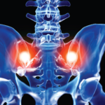
SI joint inflammation is just one cause of low back pain. Often, physical therapy rather than biologic therapy is indicated. Because rheumatologists are making this important decision based on MRI reports, MRI interpretations must be as accurate as possible.

ad goes here:advert-1
ADVERTISEMENT
SCROLL TO CONTINUE

Solutions

According to Dr. Weiss, a big contributor to this problem has been the lack of materials to familiarize radiologists with the normal maturational changes that occur in the SI joint. However, in July 2021, radiologists published an atlas of pediatric structural changes. The atlas uses the preliminary, updated JAMRIS (Juvenile Idiopathic Arthritis MRI Score) scoring system and is intended to serve as a reference for the assessment of structural lesions in patients with SI joint arthritis.2

Dr. Weiss believes these resources will help familiarize a wider audience with the appearance of the normal pediatric SI joint because “images and examples are invaluable. … I am very optimistic things are going to improve with time and awareness,” she says.

ad goes here:advert-2
ADVERTISEMENT
SCROLL TO CONTINUE

For radiologists and rheumatologists, Dr. Weiss recommends an additional resource: the educational tools created by Care Arthritis, a company focused on the development of new imaging and biomarker technologies to advance personalized medicine for patients with arthritis. Although, according to Dr. Weiss, the Care Arthritis site includes primarily adult images, the free resource also has pediatric images.


Lara C. Pullen, PhD, is a medical writer based in the Chicago area.

References

  1. Weiss PF, Brandon TG, Bohnsack J, et al. Variability in interpretation of magnetic resonance imaging of the pediatric sacroiliac joint. Arthritis Care Res (Hoboken). 2021 Jun;73(6):841–848.
  2. N Herregods, WP Maksymowych, L Jans, et al. Atlas of MRI findings of sacroiliitis in pediatric sacroiliac joints to accompany the updated preliminary OMERACT pediatric JAMRIS (Juvenile Idiopathic Arthritis MRI Score) scoring system: Part II: Structural damage lesions. Semin Arthritis Rheum. 2021 Oct;51(5):1099–1107.

Page: 1 2 3 | Single Page
Share: 

Filed under:ConditionsPediatric Conditions Tagged with:imagingjoint imagingPediatricRadiologistsacroiliac jointssacroiliitisSI joint

Related Articles

    Imaging of Axial Psoriatic Arthritis

    May 9, 2022

    The axial phenotype of psoriatic arthritis (axPsA) is an excellent example of a major controversy in rheumatology that has become the focus of attention because of the emergence of new therapies with different mechanisms of action for alleviating joint inflammation. It was first described in 1961 but, until recently, it has largely remained under the…

    Imaging in Ankylosing Spondylitis

    April 1, 2015

    MRI inflammation, fat and new bone formation in the sacroiliac joints, spine in patients with AS

    Envision Arthritis Pathology

    April 1, 2008

    MRI advances in RA and OA

    Kateryna Kon / Shutterstock.com

    How to Manage, Treat Anemia of Inflammation in Patients with Rheumatic Disease

    December 17, 2017

    Anemia is common in patients with systemic rheumatic disease, yet it may not get the attention it deserves. Anemia can result from chronic inflammation, treatment side effects or other disease factors, or it may signal an unrelated condition. Although diagnosis and treatment of anemia are sometimes challenging, clinicians must do their utmost to rigorously investigate…

  • About Us
  • Meet the Editors
  • Issue Archives
  • Contribute
  • Advertise
  • Contact Us
  • Copyright © 2025 by John Wiley & Sons, Inc. All rights reserved, including rights for text and data mining and training of artificial technologies or similar technologies. ISSN 1931-3268 (print). ISSN 1931-3209 (online).
  • DEI Statement
  • Privacy Policy
  • Terms of Use
  • Cookie Preferences