Video: Every Case Tells a Story| Webinar: ACR/CHEST ILD Guidelines in Practice

An official publication of the ACR and the ARP serving rheumatologists and rheumatology professionals

  • Conditions
    • Axial Spondyloarthritis
    • Gout and Crystalline Arthritis
    • Myositis
    • Osteoarthritis and Bone Disorders
    • Pain Syndromes
    • Pediatric Conditions
    • Psoriatic Arthritis
    • Rheumatoid Arthritis
    • Sjögren’s Disease
    • Systemic Lupus Erythematosus
    • Systemic Sclerosis
    • Vasculitis
    • Other Rheumatic Conditions
  • FocusRheum
    • ANCA-Associated Vasculitis
    • Axial Spondyloarthritis
    • Gout
    • Psoriatic Arthritis
    • Rheumatoid Arthritis
    • Systemic Lupus Erythematosus
  • Guidance
    • Clinical Criteria/Guidelines
    • Ethics
    • Legal Updates
    • Legislation & Advocacy
    • Meeting Reports
      • ACR Convergence
      • Other ACR meetings
      • EULAR/Other
    • Research Rheum
  • Drug Updates
    • Analgesics
    • Biologics/DMARDs
  • Practice Support
    • Billing/Coding
    • EMRs
    • Facility
    • Insurance
    • QA/QI
    • Technology
    • Workforce
  • Opinion
    • Patient Perspective
    • Profiles
    • Rheuminations
      • Video
    • Speak Out Rheum
  • Career
    • ACR ExamRheum
    • Awards
    • Career Development
  • ACR
    • ACR Home
    • ACR Convergence
    • ACR Guidelines
    • Journals
      • ACR Open Rheumatology
      • Arthritis & Rheumatology
      • Arthritis Care & Research
    • From the College
    • Events/CME
    • President’s Perspective
  • Search

The Brain in Lupus

Michael Lockshin, MD, and Liza Kozora, PhD  |  Issue: September 2008  |  September 1, 2008

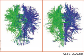
Contemporary techniques for imaging of the brain—MRI, PET, functional MRI, and diffusion tensor imaging (DTI) (see Figures 1 and 2, above)—allow the display of the anatomy of abnormal brain function and the ability to see, literally, thought in action. The images observed by these modalities do not always distinguish functional abnormalities from structural ones; also, demonstrated abnormalities may not correlate closely with cognitive function measured by formal testing. Focal breakdown of the blood–brain barrier may be critical to the localization of brain injury in diseases such as lupus, but intactness of the barrier is not easy to identify by any technology currently applicable to humans. New technologies under investigation may be more successful in defining the blood–brain barrier and may help explain the gap between the presence of autoantibodies in lupus and the occurrence focal brain changes.

Figure 2A: DTI demonstrating white track connections in a normal brain (LEFT). Figure 2B: DTI in a brain from a patient with lupus.  The quantity of tracks is less than that in a normal person (RIGHT).
Figure 2A: DTI demonstrating white track connections in a normal brain (LEFT). Figure 2B: DTI in a brain from a patient with lupus. The quantity of tracks is less than that in a normal person (RIGHT).

Lessons from the Brain

As discussed at the conference, animal models may help define mechanisms of abnormal cognition. Mice that develop clinical lupus or are exposed to lupus autoantibodies (e.g., anti-DNA, antiphospholipid, and anti-NMDA) demonstrate behavioral changes and anatomic abnormalities that parallel those of human lupus. Time-specific disruption of the blood–brain barrier, leukoagglutination, thrombosis, microvascular injury, and autoantibody-mediated cell toxicity are plausible mechanisms of brain injury demonstrable in mice, and sometimes in men.

ad goes here:advert-1
ADVERTISEMENT
SCROLL TO CONTINUE

The Mary Kirkland Center conference’s discussion of animal and human neurobiological, behavioral, and imaging information about cognition concluded that white-matter injury (interfering with brain interconnections) may be more important in lupus than is gray-matter injury, and that immunologically driven brain injury is focal for reasons not yet understood. White-matter damage, particularly in the frontal area, can parallel neuropsychologically defined global deficits of attention and executive function, and is demonstrable on MRI or DTI. Longitudinal studies suggest that cognitive dysfunction in lupus is more likely transient and reversible than permanent.

The Mary Kirkland Center conference led to the following ideas that could inform future research: Due to the wide variability of cognitive symptoms and innate capacities among patients with lupus, longitudinal study of small numbers of individual patients may be more informative than cross-sectional studies of large patient groups. Because patients’ descriptions of cognitive symptoms are clues to anatomic localization, attention to the details of responses in neuropsychological tests offers noninvasive, inexpensive opportunities for deeper study. Finally, novel experimental cognitive tests may be specifically applicable to lupus.

ad goes here:advert-2
ADVERTISEMENT
SCROLL TO CONTINUE

Table 2. Cognitive Tests for Patients with Lupus

National Adult Reading Test (NART) (to estimate IQ)*
Digit symbol substitution test
Trail-making test (part A and B)
Stroop color and word test
California verbal learning test
Rey Osterrieth complex figure test (with delayed recall)
Wechsler Adult Intelligence Scale (WAIS) III letter numbering sequencing*
Controlled oral word association test
Animal naming
Finger tapping

Page: 1 2 3 4 | Single Page
Share: 

Filed under:ConditionsEducation & TrainingResearch RheumSystemic Lupus Erythematosus Tagged with:LupusResearchSLESystemic lupus erythematosus

Related Articles

    Lupus in the Child’s Mind

    March 1, 2009

    Unique neuropsychiatric problems require a unique approach

    Pinpoint Cognitive Dysfunction in Patients with Lupus

    April 6, 2012

    More than 80% of SLE patients experience some type of neurologic manifestation during their disease course. The challenge for rheumatologists and other clinicians lies in appropriately diagnosing any cognitive dysfunctions that accompany lupus and better understanding the causes and risk factors of those dysfunctions. “Cognitive Function in SLE” was the focus of a talk at the 2011 ACR/ARHP Annual Scientific Meeting in November.

    Serological Antibody Tests in COVID-19: Test Reliability and Utility

    June 10, 2020

    Serological testing for severe acute respiratory syndrome coronavirus 2 (SARS-CoV-2) antibodies may play a critical role in the management of the worldwide health crisis. Such testing may reveal key information for epidemiology, convalescent plasma therapies and vaccine development. However, the situation is complex, and much is unknown. Although such testing may ultimately be used to…

    Stmool / shutterstock.com

    How to Avoid Cognitive Errors in Rheumatology

    March 14, 2022

    The 1999 Institute of Medicine report To Err Is Human gave a sobering depiction of the magnitude and consequences of medical error.1 The report concluded that approximately 98,000 people die in hospitals annually due to preventable medical errors. Of all the errors detailed in this report, diagnostic errors have since been determined to be the…

  • About Us
  • Meet the Editors
  • Issue Archives
  • Contribute
  • Advertise
  • Contact Us
  • Copyright © 2025 by John Wiley & Sons, Inc. All rights reserved, including rights for text and data mining and training of artificial technologies or similar technologies. ISSN 1931-3268 (print). ISSN 1931-3209 (online).
  • DEI Statement
  • Privacy Policy
  • Terms of Use
  • Cookie Preferences