Video: Every Case Tells a Story| Webinar: ACR/CHEST ILD Guidelines in Practice

An official publication of the ACR and the ARP serving rheumatologists and rheumatology professionals

  • Conditions
    • Axial Spondyloarthritis
    • Gout and Crystalline Arthritis
    • Myositis
    • Osteoarthritis and Bone Disorders
    • Pain Syndromes
    • Pediatric Conditions
    • Psoriatic Arthritis
    • Rheumatoid Arthritis
    • Sjögren’s Disease
    • Systemic Lupus Erythematosus
    • Systemic Sclerosis
    • Vasculitis
    • Other Rheumatic Conditions
  • FocusRheum
    • ANCA-Associated Vasculitis
    • Axial Spondyloarthritis
    • Gout
    • Psoriatic Arthritis
    • Rheumatoid Arthritis
    • Systemic Lupus Erythematosus
  • Guidance
    • Clinical Criteria/Guidelines
    • Ethics
    • Legal Updates
    • Legislation & Advocacy
    • Meeting Reports
      • ACR Convergence
      • Other ACR meetings
      • EULAR/Other
    • Research Rheum
  • Drug Updates
    • Analgesics
    • Biologics/DMARDs
  • Practice Support
    • Billing/Coding
    • EMRs
    • Facility
    • Insurance
    • QA/QI
    • Technology
    • Workforce
  • Opinion
    • Patient Perspective
    • Profiles
    • Rheuminations
      • Video
    • Speak Out Rheum
  • Career
    • ACR ExamRheum
    • Awards
    • Career Development
  • ACR
    • ACR Home
    • ACR Convergence
    • ACR Guidelines
    • Journals
      • ACR Open Rheumatology
      • Arthritis & Rheumatology
      • Arthritis Care & Research
    • From the College
    • Events/CME
    • President’s Perspective
  • Search

The Diagnostic View: Assess Your Rheumatology Knowledge

Jennifer L. Demertzis, MD  |  Issue: April 2015  |  April 1, 2015

Editor’s note: In this new recurring feature, we first present a series of images (this page) for your review, and then a brief discussion of the findings and diagnosis. Before you turn to the discussion, examine these images carefully and draw your own conclusions.

History

ad goes here:advert-1
ADVERTISEMENT
SCROLL TO CONTINUE

These images are of a 55-year-old man with chronic back pain.

ad goes here:advert-2
ADVERTISEMENT
SCROLL TO CONTINUE

Bilateral sacroiliac joint posteroanterior radiograph obtained at the time of the current presentation.

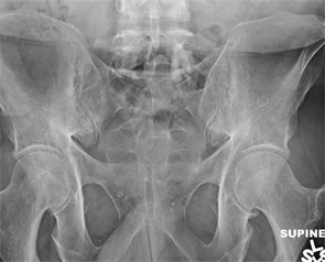
Figure 1: Bilateral sacroiliac joint posteroanterior radiograph obtained at the time of the current presentation.

Sagittal STIR image from a thoracic spine MR, obtained two years prior to the current presentation. STIR sequences are fat-suppressed, water-sensitive images, useful for detecting bone marrow and soft tissue edema.

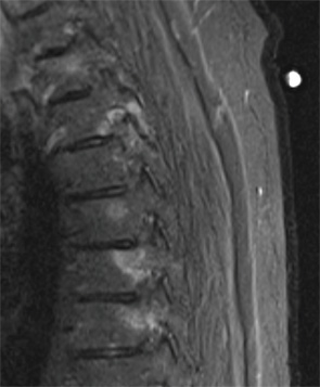
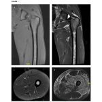
Figure 2: Sagittal STIR image from a thoracic spine MR, obtained two years prior to the current presentation. STIR sequences are fat-suppressed, water-sensitive images, useful for detecting bone marrow and soft tissue edema.

Sagittal STIR image from a lumbar spine MR, obtained two years prior to the current presentation.

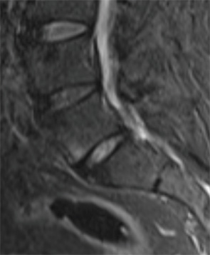
Figure 3: Sagittal STIR image from a lumbar spine MR, obtained two years prior to the current presentation.


Share: 

Filed under:Axial SpondyloarthritisConditions Tagged with:Ankylosing SpondylitisDemertzisdiagnosticimagingPainradiographrheumatologyspine MRsymmetric erosive sacroiliitis

Related Articles

    The Diagnostic View: Assess Your Rheumatology Knowledge

    March 2, 2015

    Radiograph, MR images of 54-year-old woman with right elbow pain

    Case Review: MRI Leads to Non-Rheumatic Diagnosis Surprise

    May 17, 2018

    Rheumatologists often rely on magnetic resonance imaging (MRI) in the evaluation of suspected muscular diseases. Here, we describe a case in which unexpected findings on MRI pointed to a diagnosis rarely considered as a mimicker of rheumatologic disease. The Case A 19-year-old man of Middle Eastern descent was admitted to our hospital for evaluation of…

    Imaging of Axial Psoriatic Arthritis

    May 9, 2022

    The axial phenotype of psoriatic arthritis (axPsA) is an excellent example of a major controversy in rheumatology that has become the focus of attention because of the emergence of new therapies with different mechanisms of action for alleviating joint inflammation. It was first described in 1961 but, until recently, it has largely remained under the…

    Imaging in Ankylosing Spondylitis

    April 1, 2015

    MRI inflammation, fat and new bone formation in the sacroiliac joints, spine in patients with AS

  • About Us
  • Meet the Editors
  • Issue Archives
  • Contribute
  • Advertise
  • Contact Us
  • Copyright © 2025 by John Wiley & Sons, Inc. All rights reserved, including rights for text and data mining and training of artificial technologies or similar technologies. ISSN 1931-3268 (print). ISSN 1931-3209 (online).
  • DEI Statement
  • Privacy Policy
  • Terms of Use
  • Cookie Preferences