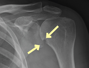
Figure 1: Left shoulder anteroposterior radiograph
Anteroposterior radiograph of the left shoulder demonstrates severe uniform narrowing of the glenohumeral joint without osteophyte formation. There are well-defined erosions of the inferior humeral head and inferior glenoid (indicated by arrows in Figure 1). Coronal T2-weighted fat-suppressed MR image demonstrates uniform full-thickness cartilage loss and erosions of both the humeral head and glenoid. The T2-weighted and T1-weighted fat-suppressed contrast-enhanced MR images confirm the presence of enhancing synovitis within the glenohumeral joint (indicated by arrows in Figure 3). Synovial tissue is seen extending into the areas of bony erosion (indicated by the circle in Figure 2. There is no bone marrow edema or osteitis. There is atrophic tendinopathy (i.e., thinning) of the rotator cuff tendons (indicated by the arrow in Figure 2).
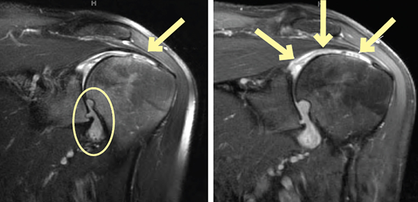
Figure 2: Left shoulder oblique coronal T2-weighted fat-suppressed MR image; Figure 3: Left shoulder oblique coronal T1-weighted fat-suppressed MR image with contrast
This constellation of radiographic and MR findings is characteristic of advanced rheumatoid arthritis in the shoulder. Although the diagnosis is confirmed on radiographs, MR may be helpful in surgical planning particularly to assess the glenoid bone stock and integrity of the rotator cuff when considering possible total shoulder arthroplasty.