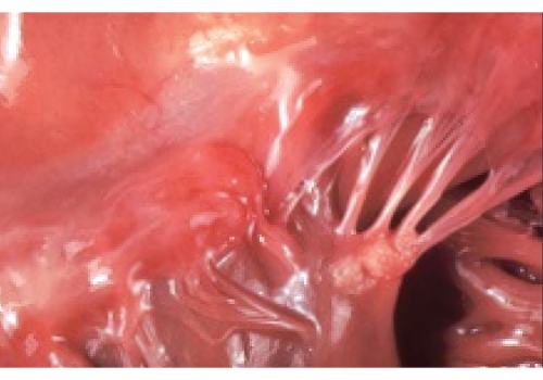
This beautifully composed photograph, without distracting features, shows Libman-Sacks endocarditis as seen at autopsy. In the days before echocardiography, clinicians knew of this lesion only by murmur, embolus or (rare) surgery for valve failure.
Dr. Matteson emphasizes just how educational and fun committee service was. “It was pure pleasure,” he says, describing interacting with the other committee members, learning about diseases, assessing the quality of slides and how they fit into the teaching program, and “of course, choosing the best and worst (spoof) slides made it all fun.”
 Alan N. Baer, MD, professor of medicine and founder of the Jerome Greene Sjögren’s Syndrome Clinic at Johns Hopkins University School of Medical, Baltimore, ushered the image collection into its current form—the ACR Image Library. Serving as committee chair from 2008–10, he worked with the committee to convert the entire collection of more than 1,000 slides to an online image bank, making the collection available to ACR members as a membership benefit.
Updating the Image Library to an online format has allowed for the inclusion of other technological updates, such as videos. “With the online format, we were able to expand our inclusion criteria for submissions,” says Dr. Baer. “Thus, we could now take videos and case series—multiple images devoted to one case.”
‘The Image Library is a great teaching tool for trainees & a repository of images for conditions/complications that may become increasingly rare as better treatments become available for our diseases.’ —Dr. Baer
One advancement that Dr. Baer was not able to get implemented was the idea of making each image submission a scholarly product that could be listed by the submitter on their curriculum vitae to count toward professional advancement (e.g., promotion, fellowships). “This idea has never been adopted,” he says. “The submissions remain anonymous to the users of the image bank.”
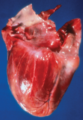
This gross specimen demonstrates a relatively normal coronary artery on the left and “beading” of the coronary artery on the right, secondary to segmental necrotizing arteritis.
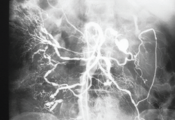
A selective angiogram of the superior mesenteric artery shows multiple saccular aneurysms that vary in size and shape. Note the irregularity of the vascular lumens with areas of stenosis and dilation.
 The most recent committee chair, Chris Collins, MD, medical director for Aurinia Pharmaceuticals, Rockville, Md., and formerly an associate professor of medicine at Georgetown University Medical Center, Washington, D.C., continues the long tradition of expanding the Image Library to keep up with the ever-changing technology.
All images, from the 191 original slides to the thousands currently in the collection to the new ones added each year, will “remain in perpetuity to provide an educational reservoir of what we see in clinical practice,” says Dr. Collins.