Video: Every Case Tells a Story| Webinar: ACR/CHEST ILD Guidelines in Practice

An official publication of the ACR and the ARP serving rheumatologists and rheumatology professionals

  • Conditions
    • Axial Spondyloarthritis
    • Gout and Crystalline Arthritis
    • Myositis
    • Osteoarthritis and Bone Disorders
    • Pain Syndromes
    • Pediatric Conditions
    • Psoriatic Arthritis
    • Rheumatoid Arthritis
    • Sjögren’s Disease
    • Systemic Lupus Erythematosus
    • Systemic Sclerosis
    • Vasculitis
    • Other Rheumatic Conditions
  • FocusRheum
    • ANCA-Associated Vasculitis
    • Axial Spondyloarthritis
    • Gout
    • Psoriatic Arthritis
    • Rheumatoid Arthritis
    • Systemic Lupus Erythematosus
  • Guidance
    • Clinical Criteria/Guidelines
    • Ethics
    • Legal Updates
    • Legislation & Advocacy
    • Meeting Reports
      • ACR Convergence
      • Other ACR meetings
      • EULAR/Other
    • Research Rheum
  • Drug Updates
    • Analgesics
    • Biologics/DMARDs
  • Practice Support
    • Billing/Coding
    • EMRs
    • Facility
    • Insurance
    • QA/QI
    • Technology
    • Workforce
  • Opinion
    • Patient Perspective
    • Profiles
    • Rheuminations
      • Video
    • Speak Out Rheum
  • Career
    • ACR ExamRheum
    • Awards
    • Career Development
  • ACR
    • ACR Home
    • ACR Convergence
    • ACR Guidelines
    • Journals
      • ACR Open Rheumatology
      • Arthritis & Rheumatology
      • Arthritis Care & Research
    • From the College
    • Events/CME
    • President’s Perspective
  • Search

These 3 Tough Cases from the 2018 Thieves Market Underscore Need for Clinical Diligence

Thomas R. Collins  |  Issue: February 2019  |  February 17, 2019

Case 2

Paras Karmacharya, MBBS, a rheum­atology fellow at Mayo Clinic in Rochester, Minn., presented a case involving a 55-year-old man with metastatic melanoma on checkpoint-inhibitor therapy (nivolumab) who had had worsening proximal muscle weakness over seven weeks, with drooping eyelids, disturbances to his vision, fatigue and weight loss.

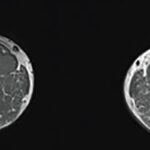
He was taking prednisone, along with vitamin D3, omeprazole and temezepam. The main lab finding was elevated muscle enzymes. He had streaky intramuscular edema on magnetic resonance imaging (MRI), and diffuse myopathy that most severely affected his bulbar, axial and proximal upper limb muscles on electro­myography. A muscle biopsy suggested necrotizing myopathy.

ad goes here:advert-1
ADVERTISEMENT
SCROLL TO CONTINUE

The diagnosis? PD-1 inhibitor-associated necrotizing myopathy.

“One salient feature that distinguishes this is the ocular muscle involve­ment seen in this case,” said Dr. Karmacharya.

ad goes here:advert-2
ADVERTISEMENT
SCROLL TO CONTINUE

The patient’s nivolumab was discontinued. He was started on methylprednisone, but he developed persistent dysphagia and was switched to intravenous immunoglobulin therapy. After that, his symptoms improved.

Neuromuscular complications due to checkpoint inhibitor treatment are rare, but can prove life-threatening. Prompt recognition and treatment can help outcomes, but Dr. Karmacharya also said the “long-term outcome for this rare man­ifestation is unknown, and it is unclear if [patients] require a steroid-sparing agent.”

Case 3

Priyanka Iyer, MBBS, MPH, a rheumatology fellow at the University of Iowa, Iowa City, presented the case of a 53-year-old man who had undergone a liver transplant for alcoholic cirrhosis and was treated with cyclosporine and acetaminophen. He presented with bilateral knee effusion, pleural weakness and myalgias.

His labs revealed signs of anemia, an elevated erythrocyte sedimentation rate, and bone-specific alkaline phosphatase. An MRI showed small, bilateral knee effusion and bone marrow edema. A bone scan was notable for symmetric uptake in both knees and both ankles.

“Inflammatory arthritis was on top of our list,” Dr. Iyer said. “This is what we see most commonly in the clinic. But his presentation was somewhat odd.” He had severe pain but didn’t have many signs of inflammation, she said.

One by one, clinicians ruled out septic arthritis, myositis, avascular necrosis and other potential diagnoses. Then, she said, clinicians thought about his symptom timeline.

“We wondered if something we [did] post-transplant could be the cause of his complaints,” she said. Based on his elevated bone-specific alkaline phosphatase level, abnormal MRI and bone scan, they settled on a diagnosis of calcineurin inhibitor pain syndrome.

Cyclosporine and tacrolimus are the most common culprits, with symmetric lower extremity arthralgias (and sometimes long bone pain) as important signals. The pain is brought on by the calcineurin inhibitor when the drug causes vascular changes, leading to disturbance of bone perfusion and permeability, intraosseous vasoconstriction, bone marrow edema and pain. Consider calcium channel blockers as an adjunct treatment, Dr. Iyer said.

In this case, the transplant team tried alternate therapies such as mycophenolate mofetil and sirolimus, but the patient didn’t tolerate them well. He was therefore restarted on a lower dose of cyclosporine with careful drug trough monitoring, with nifedipine and pregabalin finally added. The man’s symptoms improved, but it took months, Dr. Iyer said.

Although calcineurin inhibitor pain syndrome is rare, it is important to be aware of it. “It may affect up to one in 20 patients on calcineurin inhibitors,” Dr. Iyer said. 

Page: 1 2 3 | Single Page
Share: 

Filed under:ConditionsMeeting ReportsSystemic Lupus Erythematosus Tagged with:2018 ACR/ARHP Annual MeetingCancercase reportCheckpoint InhibitorscyclosporineImmunoglobinleprosynifedipinepregabalintacrolimus

Related Articles

    New Tools for Myositis Diagnosis, Classification & Management

    April 15, 2019

    CHICAGO—At Hot Topics in Myositis, a session at the 2018 ACR/ARHP Annual Meeting, three experts discussed new classification criteria for idiopathic inflammatory myopathies (IIM) and offered practical primers on overlap myositis conditions and inclusion body myositis (IBM). New Myositis Classification Criteria After a 10-year development process, the new EULAR/ACR Classification Criteria for Adult and Juvenile…

    A Primer on Imaging in Myositis

    November 9, 2022

    In medicine, as in advertising, pictures can be worth a thousand words. From arthritis to vasculitis, imaging studies have been variably employed to aid in the diagnosis, treatment, risk stratification and prognostication of patients with rheumatic and musculoskeletal disorders. The same holds true with the idiopathic inflammatory myopathies (IIM), in which the clinical utility is…

    Diagnosis: Myopathy

    July 1, 2009

    Presentation and evaluation of metabolic causes

    A Duet of Bone and the Immune System

    July 12, 2011

    Examining emerging perspectives in osteoimmunology

  • About Us
  • Meet the Editors
  • Issue Archives
  • Contribute
  • Advertise
  • Contact Us
  • Copyright © 2025 by John Wiley & Sons, Inc. All rights reserved, including rights for text and data mining and training of artificial technologies or similar technologies. ISSN 1931-3268 (print). ISSN 1931-3209 (online).
  • DEI Statement
  • Privacy Policy
  • Terms of Use
  • Cookie Preferences