Video: Every Case Tells a Story| Webinar: ACR/CHEST ILD Guidelines in Practice

An official publication of the ACR and the ARP serving rheumatologists and rheumatology professionals

  • Conditions
    • Axial Spondyloarthritis
    • Gout and Crystalline Arthritis
    • Myositis
    • Osteoarthritis and Bone Disorders
    • Pain Syndromes
    • Pediatric Conditions
    • Psoriatic Arthritis
    • Rheumatoid Arthritis
    • Sjögren’s Disease
    • Systemic Lupus Erythematosus
    • Systemic Sclerosis
    • Vasculitis
    • Other Rheumatic Conditions
  • FocusRheum
    • ANCA-Associated Vasculitis
    • Axial Spondyloarthritis
    • Gout
    • Psoriatic Arthritis
    • Rheumatoid Arthritis
    • Systemic Lupus Erythematosus
  • Guidance
    • Clinical Criteria/Guidelines
    • Ethics
    • Legal Updates
    • Legislation & Advocacy
    • Meeting Reports
      • ACR Convergence
      • Other ACR meetings
      • EULAR/Other
    • Research Rheum
  • Drug Updates
    • Analgesics
    • Biologics/DMARDs
  • Practice Support
    • Billing/Coding
    • EMRs
    • Facility
    • Insurance
    • QA/QI
    • Technology
    • Workforce
  • Opinion
    • Patient Perspective
    • Profiles
    • Rheuminations
      • Video
    • Speak Out Rheum
  • Career
    • ACR ExamRheum
    • Awards
    • Career Development
  • ACR
    • ACR Home
    • ACR Convergence
    • ACR Guidelines
    • Journals
      • ACR Open Rheumatology
      • Arthritis & Rheumatology
      • Arthritis Care & Research
    • From the College
    • Events/CME
    • President’s Perspective
  • Search

Ultrasound Image Review: A 30-Year-Old Woman with Left Foot Pain

Eugene Kissin, MD, RhMSUS, & Catherine Bakewell, MD, RhMSUS  |  Issue: May 2019  |  May 17, 2019

Presentation

A 30-year-old woman presented to her rheumatologist for left foot pain of three weeks’ duration. She was followed for systemic lupus erythematosus manifesting in arthritis and hemolytic anemia, as well as anti-nuclear antibody and Smith antibody positivity, and was treated with hydroxychloroquine and prednisone in the 2.5–10 mg per day range. She was symptom free for six months prior to the development of her foot pain and had tapered off prednisone six weeks prior to her visit. Every day for the previous month, she had walked a mile to the hospital for her sick child. Her foot pain was much worse with weight bearing, but she had no other symptoms of disease. Her examination revealed normal vital signs and localized tenderness over the third metatarsal.

The Ultrasound

Panel “a” in the figure above shows a longitudinal ultrasound image of the dorsal aspect of the third metatarsal, with hazy, hyperechoic material over the metatarsal shaft, outlined by an anechoic lining. This appearance is produced by periosteal elevation and edema. The arrow head points to synovial tissue of the metatarsal joint, which was neither tender to sonopalpation, nor hyperemic on Doppler imaging.

ad goes here:advert-1
ADVERTISEMENT
SCROLL TO CONTINUE

Panels “b” and “c” demonstrate hyperemia surrounding the periosteum on longitudinal and transverse Doppler imaging, but not the joint synovium.

Ultrasound Image Review: A 30-Year-Old Woman with Left Foot Pain
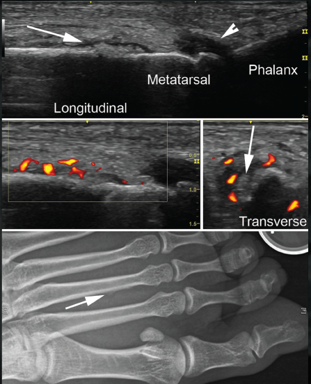
Panel “d” depicts a radiograph of the foot, with an arrow pointing to the barely visible elevated periosteum, corresponding to the region of stress fracture.

ad goes here:advert-2
ADVERTISEMENT
SCROLL TO CONTINUE

Treatment & Discussion

The patient was treated with a walking boot, and her symptoms resolved. Ultrasound can demonstrate superficial bone lesions involving the periosteum, including stress fractures, and may be able to detect such lesions more easily than pain film radiography, although this question has not yet been systematically evaluated.1

One study published last year did find that emergency medicine residents were better able to identify long bone fractures using ultrasound than with X-ray (accurate identification mean proportion 0.89 vs. 0.75, respectively [P<0.001]). This difference was more pronounced as the level of ultrasound and emergency department experience increased.2

Stress fractures are more likely to occur in patients with rheumatic conditions, not only due to a predisposition to osteoporosis from the condition itself, but often due to concomitant treatment with glucocorticoids. Stress fractures of the tibia, metatarsals and fibula are the most frequently reported.3


The authors are mentors for USSONAR, a society of rheumatologists, physicians and health professionals working to promote the use of musculoskeletal ultra­sonography to advance the care of patients with rheumatic diseases. Learn more.

Discover

The ACR offers an RhMSUS certification program. Learn more.

Page: 1 2 | Single Page
Share: 

Filed under:Conditions Tagged with:FracturesGlucocorticoidsstress fractureUltrasound

Related Articles

    Osteoporosis Experts Discuss Bisphosphonate Holidays

    November 24, 2020

    ACR CONVERGENCE 2020—Bisphosphonates are an important treatment for millions of older Americans with osteoporosis because the drugs inhibit osteoclastic bone resorption to reduce the risk of painful, debilitating fractures.1 More than 20 years ago, data emerged that bisphosphonates have a long terminal half-life.2 So after years of therapy, could some patients take a drug holiday?…

    Foot Pain a Burden for Rheumatology Patients

    January 13, 2012

    Researchers say it may even correlate with back, hip, and knee pain

    Podiatry for Rheumatologists: Treating Patients’ Foot Complaints

    April 1, 2015

    Tips for diagnosing, managing hallux rigidus, metatarsalgia, neuroma and rear foot problems in rheumatology patients

    Changing Our Thinking on Osteoarthritis

    March 1, 2010

    It’s time we changed our thinking on osteoarthritis

  • About Us
  • Meet the Editors
  • Issue Archives
  • Contribute
  • Advertise
  • Contact Us
  • Copyright © 2025 by John Wiley & Sons, Inc. All rights reserved, including rights for text and data mining and training of artificial technologies or similar technologies. ISSN 1931-3268 (print). ISSN 1931-3209 (online).
  • DEI Statement
  • Privacy Policy
  • Terms of Use
  • Cookie Preferences