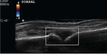
Video: Knock on Wood| Webinar: ACR/CHEST ILD Guidelines in Practice
fa-facebookfa-linkedinfa-youtube-playfa-rss

An official publication of the ACR and the ARP serving rheumatologists and rheumatology professionals

  • Conditions
    • Axial Spondyloarthritis
    • Gout and Crystalline Arthritis
    • Myositis
    • Osteoarthritis and Bone Disorders
    • Pain Syndromes
    • Pediatric Conditions
    • Psoriatic Arthritis
    • Rheumatoid Arthritis
    • Sjögren’s Disease
    • Systemic Lupus Erythematosus
    • Systemic Sclerosis
    • Vasculitis
    • Other Rheumatic Conditions
  • FocusRheum
    • ANCA-Associated Vasculitis
    • Axial Spondyloarthritis
    • Gout
    • Lupus Nephritis
    • Psoriatic Arthritis
    • Rheumatoid Arthritis
    • Systemic Lupus Erythematosus
  • Guidance
    • Clinical Criteria/Guidelines
    • Ethics
    • Legal Updates
    • Legislation & Advocacy
    • Meeting Reports
      • ACR Convergence
      • Other ACR meetings
      • EULAR/Other
    • Research Rheum
  • Drug Updates
    • Analgesics
    • Biologics/DMARDs
  • Practice Support
    • Billing/Coding
    • EMRs
    • Facility
    • Insurance
    • QA/QI
    • Technology
    • Workforce
  • Opinion
    • Patient Perspective
    • Profiles
    • Rheuminations
      • Video
    • Speak Out Rheum
  • Career
    • ACR ExamRheum
    • Awards
    • Career Development
  • ACR
    • ACR Home
    • ACR Convergence
    • ACR Guidelines
    • Journals
      • ACR Open Rheumatology
      • Arthritis & Rheumatology
      • Arthritis Care & Research
    • From the College
    • Events/CME
    • President’s Perspective
  • Search

Ultrasound Training Tips & Pitfalls

Eugene Kissin, MD, RhMSUS; Paul J. DeMarco, MD, FACP, FACR, RhMSUS; Daniel G. Malone, MD, RMSK; Catherine Bakewell, MD, RhMSUS; Karina Torralba, MD, FACR, CCD, RhMSUS; Rany al Haj, MD; Narandra Bethina, MD, RhMSUS; Clara Lin, MD, RhMSUS; Amy M. Evangelisto, MD, RhMSUS; Jay Higgs, MD, RhMSUS; & Midori Jane Nishio, MD, RhMSUS  |  Issue: November 2018  |  November 19, 2018

Ankle

The Doppler View

Figure 3
The Doppler View

Half of the participants stumbled in visualizing the posterior tibial tendon (PTT) insertion. The course of the PTT in the ankle and foot is as follows: around the posterior portion of the medial malleolus, then on the cranial surface of the sustentaculum tali, then inferior to the calcaneo-
navicular (i.e., spring) ligament. Finally, as it gets to the navicular, about half of its fibers attach to the navicular tuberosity (infero-medial surface of the navicular) and the medial cuneiform. Some recurrent fibers connect to the sustentaculum tali, and the remaining fibers continue to the plantar surface of the foot. Due to this unusual course, when imaging the PTT in its longitudinal view in the medial foot, the sound beam must be angled slightly cephalad to minimize anisotropy.4

A few fellows misidentified calcaneal erosion as osteophytes due to failure to correlate longitudinal with transverse images and construct a three-dimensional image of the Achilles insertion in their minds. Erosions are potholes in the bone, and osteophytes are jagged extrusions from the bone. These two will be confused only if the sonographer cannot determine where the normal bone surface is, and this mistake could lead to substantial diagnostic and treatment errors (see Figure 1). At this moment, the student tends to feel like a heel (sic, you know who).

ad goes here:advert-1
ADVERTISEMENT
SCROLL TO CONTINUE

1st Metatarsalphalangeal Joint

The synovial reflection for the dorsal aspect of this joint is superficial and easily compressed by even light transducer pressure. Many students scan the first MTP without floating the transducer, a technique in which a thick layer of gel is maintained between the surface of the skin and the transducer face to minimize tissue pressure. The small effusion the model had was missed by the students who did not employ the floating technique.

The other difficulty for students included obtaining a longitudinal view of the plantar/volar metatarsal head due to shadowing from the sesamoid bones, and some resulting confusion of the sesamoid bones with the metatarsal head. The metatarsal head is deeper and has a layer of hyaline cartilage that the sesamoids do not. Often, the transducer frequency must be lowered for this view to penetrate the thick plantar soft tissues.

ad goes here:advert-2
ADVERTISEMENT
SCROLL TO CONTINUE

The Bottom Line

Despite a formal standardized training program, considerable variability in participant performance on written and practical examinations exists. We believe our final exam results not only evaluate competence, but also help fill in skill and knowledge gaps among the trainees and align their confidence to their competence. The ACR’s Musculoskeletal Ultrasound Certification in Rheumatology (RhMSUS) certification examination in musculoskeletal ultrasound also serves this important purpose: to let the practitioner know whether their ultrasound knowledge level meets acceptable standards. This is a very valuable service to the rheumatology community and our patients.

Although RhMSUS does not include a practical examination, there is a very high correlation between the participant scores on the written and practical portions of other examinations.5  

Page: 1 2 3 4 5 | Single Page
Share: 

Filed under:CertificationEducation & TrainingEducation & Training Tagged with:RhMSUSUltrasound

Related Articles
    A transverse view of the ulnar groove in full elbow extension. The red arrow indicates the advancing edge of the MHTr.

    Recurrent Medial Elbow Pain Following Successful Tommy John Surgery

    August 12, 2020

    A 27-year-old, left-handed man was referred to our ultrasound clinic for left elbow pain. History The patient had been a pitcher on a Minor League Baseball team. Two years before, he developed sudden, severe medial elbow pain while pitching in a game. The pain was associated with some tingling down the left medial forearm. The…

    Musculoskeletal Ultrasound: A Valuable Tool for Diagnosing Rheumatic Illnesses

    Musculoskeletal Ultrasound: A Valuable Tool for Diagnosing Rheumatic Illnesses

    October 15, 2015

    Musculoskeletal (MSK) ultrasound is a valuable imaging modality for the practicing rheumatologist and provides an efficient tool with high diagnostic value in the evaluation of patients with musculoskeletal complaints. The use of MSK ultrasound has evolved in the U.S. due to the emergence of less-expensive, portable ultrasound units, which provide high-quality gray-scale and power Doppler…

    Ortho Angle

    August 1, 2009

    Where rheumatology and orthopedics meet

    Tips for Implementing Ultrasound Training in Rheumatology Fellowships

    February 13, 2020

    ATLANTA—Point-of-care ultrasound education mainly has occurred at the undergraduate level at U.S. medical schools, but rheumatology fellowship training programs are rapidly catching up and integrating it into their curricula, according to two program directors who reviewed the state of rheumatology ultrasound education, including potential barriers to its implementation, on Nov. 12 at the 2019 ACR/ARP…

  • About Us
  • Meet the Editors
  • Issue Archives
  • Contribute
  • Advertise
  • Contact Us
fa-facebookfa-linkedinfa-youtube-playfa-rss
  • Copyright © 2025 by John Wiley & Sons, Inc. All rights reserved, including rights for text and data mining and training of artificial technologies or similar technologies. ISSN 1931-3268 (print). ISSN 1931-3209 (online).
  • DEI Statement
  • Privacy Policy
  • Terms of Use
  • Cookie Preferences