Video: Every Case Tells a Story| Webinar: ACR/CHEST ILD Guidelines in Practice

An official publication of the ACR and the ARP serving rheumatologists and rheumatology professionals

  • Conditions
    • Axial Spondyloarthritis
    • Gout and Crystalline Arthritis
    • Myositis
    • Osteoarthritis and Bone Disorders
    • Pain Syndromes
    • Pediatric Conditions
    • Psoriatic Arthritis
    • Rheumatoid Arthritis
    • Sjögren’s Disease
    • Systemic Lupus Erythematosus
    • Systemic Sclerosis
    • Vasculitis
    • Other Rheumatic Conditions
  • FocusRheum
    • ANCA-Associated Vasculitis
    • Axial Spondyloarthritis
    • Gout
    • Psoriatic Arthritis
    • Rheumatoid Arthritis
    • Systemic Lupus Erythematosus
  • Guidance
    • Clinical Criteria/Guidelines
    • Ethics
    • Legal Updates
    • Legislation & Advocacy
    • Meeting Reports
      • ACR Convergence
      • Other ACR meetings
      • EULAR/Other
    • Research Rheum
  • Drug Updates
    • Analgesics
    • Biologics/DMARDs
  • Practice Support
    • Billing/Coding
    • EMRs
    • Facility
    • Insurance
    • QA/QI
    • Technology
    • Workforce
  • Opinion
    • Patient Perspective
    • Profiles
    • Rheuminations
      • Video
    • Speak Out Rheum
  • Career
    • ACR ExamRheum
    • Awards
    • Career Development
  • ACR
    • ACR Home
    • ACR Convergence
    • ACR Guidelines
    • Journals
      • ACR Open Rheumatology
      • Arthritis & Rheumatology
      • Arthritis Care & Research
    • From the College
    • Events/CME
    • President’s Perspective
  • Search

Updates on Giant Cell Arteritis

Susan Bernstein  |  Issue: March 2018  |  March 19, 2018

Patients with upper-extremity limb claudication may be younger than others with GCA, who have more typical cranial manifestations.9 Their diagnosis may be delayed because they lack the more common signs of GCA, said Dr. Kermani. “Maybe physicians don’t recognize it or think about it early enough. Inflammation may be in different segments, so they tend to have less vision loss,” she said. These GCA patients may have a negative temporal artery biopsy. “So if you suspect this diagnosis, large-vessel imaging is really the way to proceed in terms of your diagnostic evaluation.”

Although GCA patients should be screened for aortic aneurysms, a late-stage complication, there is no consensus on the type of imaging modality or frequency and timing of screening in these patients, she said. GCA patients are also at increased risk for vertebrobasilar stroke, myocardial infarction and venous thromboembolism, so GCA screening may be considered in older patients who present with these vascular events.10

ad goes here:advert-1
ADVERTISEMENT
SCROLL TO CONTINUE

Glucocorticoids have been the mainstay of GCA therapy in the past, and “prompt treatment in anyone with suspected GCA is essential to prevent vision loss. In patients where vision loss does occur, prompt institution of glucocorticoids can prevent contralateral eye involvement or progression of their visual deficit,” said Dr. Kermani. After a starting dose of 40–60 mg/day for one month, if symptoms resolve and biomarkers normalize, patients may taper their glucocorticoid dose. Patients with vision loss or ischemic complications such as limb claudication may need pulse steroids, she added. Aspirin is not routinely recommended.

Tocilizumab may help GCA patients lower their overall steroid dose and decrease relapse risk, according to the results of a Phase III, randomized clinical trial.11 Other potential therapies now in clinical trials include abatacept and ustekinumab, but these are not yet approved for GCA, she said.12,13

ad goes here:advert-2
ADVERTISEMENT
SCROLL TO CONTINUE

Imaging Strengths & Limitations

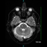
Rheumatologists must select the right imaging modality at different points in the GCA disease process, said Peter C. Grayson, MD, MSc, Principal Investigator of the translational research program in vasculitis at the National Institutes of Arthritis and Musculoskeletal and Skin Diseases (NIAMS) of the National Institutes of Health (NIH). Ultrasound of extracranial vessels to diagnose GCA has many advantages, he said.14 It is relatively cheap, non-invasive, involves no radiation and can be performed at bedside.

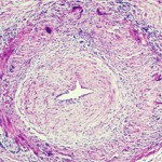
“It’s a dynamic form of imaging. You can see how blood is flowing through the arteries and see signs of turbulence,” said Dr. Grayson. “The other thing you can see in ultrasound is the wall of the artery and the morphology of it. Is this imaging finding telling me that inflammation is going on in this vessel wall? What you will see, particularly in the cross-sectional view, is you can see this hypo-echoic rim around the lumen of the artery, which is what we call the halo sign. This tells me that there is likely this edematous, thickened arterial wall,” a sign of potential arteritis.

Page: 1 2 3 4 5 6 | Single Page
Share: 

Filed under:Meeting ReportsVasculitis Tagged with:ACR/ARHP Annual Meetinggiant cell arteritis (GCA)

Related Articles

    Two Inflammatory Conditions—Polymyalgia Rheumatica and Giant Cell Arteritis—Share Clinical Connection

    March 1, 2013

    Polymyalgia rheumatica (PMR) and giant cell arteritis (GCA) have common clinical and epidemiologic links, but they need not occur synchronously

    Giant Cell Arteritis Challenging to Diagnose, Manage

    March 1, 2015

    Common form of primary vasculitis difficult to identify, treat, but latest research suggests potential new therapeutic targets

    Case Report: Giant Cell Arteritis-Related Stroke

    September 10, 2023

    Thromboembolic events are major contributors to the morbidity and mortality of patients with giant cell arteritis (GCA), but little is known about how GCA may increase the risk of ischemic strokes. GCA-related stroke is described as an ischemic cerebral infarct occurring within three to four weeks of GCA diagnosis and treatment. It occurs in 3–7%…

    Research in Temporal Arteritis Suggests Link with Infection, Autoimmune Disease

    November 16, 2015

    Temporal arteritis was first described by Sir Jonathan Hutchinson in 1890 in an elderly retired gentleman’s servant who developed red, painful streaks on his temples and was found to have bilaterally swollen temporal arteries with feeble pulses.1 Sir Hutchinson disputed the suggestion that the red streaks were caused by the man’s hat and, instead, called…

  • About Us
  • Meet the Editors
  • Issue Archives
  • Contribute
  • Advertise
  • Contact Us
  • Copyright © 2025 by John Wiley & Sons, Inc. All rights reserved, including rights for text and data mining and training of artificial technologies or similar technologies. ISSN 1931-3268 (print). ISSN 1931-3209 (online).
  • DEI Statement
  • Privacy Policy
  • Terms of Use
  • Cookie Preferences