Video: Every Case Tells a Story| Webinar: ACR/CHEST ILD Guidelines in Practice

An official publication of the ACR and the ARP serving rheumatologists and rheumatology professionals

  • Conditions
    • Axial Spondyloarthritis
    • Gout and Crystalline Arthritis
    • Myositis
    • Osteoarthritis and Bone Disorders
    • Pain Syndromes
    • Pediatric Conditions
    • Psoriatic Arthritis
    • Rheumatoid Arthritis
    • Sjögren’s Disease
    • Systemic Lupus Erythematosus
    • Systemic Sclerosis
    • Vasculitis
    • Other Rheumatic Conditions
  • FocusRheum
    • ANCA-Associated Vasculitis
    • Axial Spondyloarthritis
    • Gout
    • Psoriatic Arthritis
    • Rheumatoid Arthritis
    • Systemic Lupus Erythematosus
  • Guidance
    • Clinical Criteria/Guidelines
    • Ethics
    • Legal Updates
    • Legislation & Advocacy
    • Meeting Reports
      • ACR Convergence
      • Other ACR meetings
      • EULAR/Other
    • Research Rheum
  • Drug Updates
    • Analgesics
    • Biologics/DMARDs
  • Practice Support
    • Billing/Coding
    • EMRs
    • Facility
    • Insurance
    • QA/QI
    • Technology
    • Workforce
  • Opinion
    • Patient Perspective
    • Profiles
    • Rheuminations
      • Video
    • Speak Out Rheum
  • Career
    • ACR ExamRheum
    • Awards
    • Career Development
  • ACR
    • ACR Home
    • ACR Convergence
    • ACR Guidelines
    • Journals
      • ACR Open Rheumatology
      • Arthritis & Rheumatology
      • Arthritis Care & Research
    • From the College
    • Events/CME
    • President’s Perspective
  • Search

Using Ultrasound to Diagnose Carpal Tunnel Syndrome

Mark H. Greenberg, MD, RMSK, RhMSUS, Julian Greer, & James W. Fant Jr., MD  |  Issue: April 2018  |  April 26, 2018

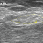
Sonography offers both qualitative and quantitative criteria to support the presence of CTS. Qualitatively, the median nerve may become hypoechoic and lose normal nerve echotexture.1 There may be swelling of the nerve proximal to the compression, and flattening of the nerve within a compression area. The abrupt flattening of the nerve is termed the “notch sign.”8

Various numeric sonographic criteria for CTS have been proposed, including direct measurement of CSA of the enlarged median nerve, comparison of CSA of the enlarged median nerve to the value in the forearm (i.e., the wrist-to-forearm ratio, or WFR), and the degree of palmar bowing of the flexor retinaculum.9-11

ad goes here:advert-1
ADVERTISEMENT
SCROLL TO CONTINUE

But how accurate is US in diagnosing CTS? A 2012 evidence-based guideline reviewed the four highest-quality studies up until May 2011 and determined the sensitivity of CTS diagnosis by US ranged from 65–97%, with accuracy ranging from 79–97%.5,12 These studies relied on median nerve CSA with the cut-off ranging from 8.5–10 mm2.

The conclusion: Based on Class I and Class II evidence, US measurement of median nerve CSA at the wrist is accurate and may be offered as a diagnostic test for CTS (Level A).12

ad goes here:advert-2
ADVERTISEMENT
SCROLL TO CONTINUE

What about Electrophysiological Studies?

Comparing US to EPS for CTS diagnosis may not be fair, because US delineates nerve structure and EPS looks at nerve function. In multiple studies, US and EPS featured fairly good correlation, although US correlated less well for mild CTS.13-16 We need further studies, though, before an abnormal US carries the same weight as an abnormal EPS in confirming CTS diagnosis.

What about patients with clinical CTS and negative EPS? As previously described, one study found completely normal EPS in 10–15% of cases of clinical CTS, and another study found 25% of CTS patients had normal EPS.6,17 A third study looked at 319 patients with a clinical diagnosis of CTS in whom 59 had normal EPS. US was performed on those 59 patients and compared with normal controls. US confirmed CTS in 18 of the EPS-negative 59 patients (i.e., about 30%).

The conclusion: US may be useful in diagnosing CTS in EPS-negative patients.18

How Do Orthopedists View US Diagnosis?

Not with any consensus. An evidence-based Level III meta-analysis of 19 articles (with a total of 3,131 wrists) published in an orthopedic journal in 2010 described US sensitivity and specificity of 77.6% and 86.8%, respectively. The authors suggested US, although not as sensitive or specific as EPS, may be a feasible alternative to EPS as a first-line confirmatory test.19 However, the February 2016 American Academy of Orthopaedic Surgeons’ CTS management guidelines support not routinely using US for the diagnosis of CTS due to limited evidence.20

Page: 1 2 3 4 5 6 | Single Page
Share: 

Filed under:ConditionsSoft Tissue Pain Tagged with:Carpal Tunnel Syndromecase reportelectrophysiological studiesUltrasound

Related Articles
    Musculoskeletal Ultrasound: A Valuable Tool for Diagnosing Rheumatic Illnesses

    Musculoskeletal Ultrasound: A Valuable Tool for Diagnosing Rheumatic Illnesses

    October 15, 2015

    Musculoskeletal (MSK) ultrasound is a valuable imaging modality for the practicing rheumatologist and provides an efficient tool with high diagnostic value in the evaluation of patients with musculoskeletal complaints. The use of MSK ultrasound has evolved in the U.S. due to the emergence of less-expensive, portable ultrasound units, which provide high-quality gray-scale and power Doppler…

    Patients with Non-Classic Carpal Tunnel Syndrome Can Benefit from Surgery

    September 19, 2014

    Patients with extra-median spread of CTS symptoms had similar post-surgical outcomes as patients with classic CTS

    Patients with Parkinson’s Disease Are at Risk for Carpal Tunnel Syndrome

    November 21, 2016

    Patients with Parkinson’s disease may be at risk of carpal tunnel syndrome (CTS). New research examined why patients with Parkinson’s have a higher incidence of CTS than that of the general population. The study found that those patients who underwent subthalamic nucleus deep brain stimulation to monitor them for neuropathy may develop CTS…

    Right: The same view as 2A, with the common peroneal nerve outlined in yellow with a cross-sectional area of 21 mm2.

    Case Report: Ultrasound Reveals Cause of Post-Arthroplasty Knee Pain

    April 15, 2020

    A 65-year-old woman was referred by an orthopedist to a rheumatologist for left knee pain. Previously, in 2014, she underwent left total knee arthroplasty (TKA) for severe osteoarthritis in a different institution. Following the procedure, she experienced severe chronic anterolateral knee pain at rest, exacerbated by walking. Because she was rendered wheelchair bound and required…

  • About Us
  • Meet the Editors
  • Issue Archives
  • Contribute
  • Advertise
  • Contact Us
  • Copyright © 2025 by John Wiley & Sons, Inc. All rights reserved, including rights for text and data mining and training of artificial technologies or similar technologies. ISSN 1931-3268 (print). ISSN 1931-3209 (online).
  • DEI Statement
  • Privacy Policy
  • Terms of Use
  • Cookie Preferences