Foot osteoarthritis (OA) is increasingly recognized as a major contributor to the overall pain and burden of OA, affecting approximately one in six adults older than 50 and greatly affecting physical function and quality of life (QoL).1 In the foot, the first metatarsophalangeal joint (great toe) is the most common reported site for symptomatic OA, followed by several joints of the midfoot.1

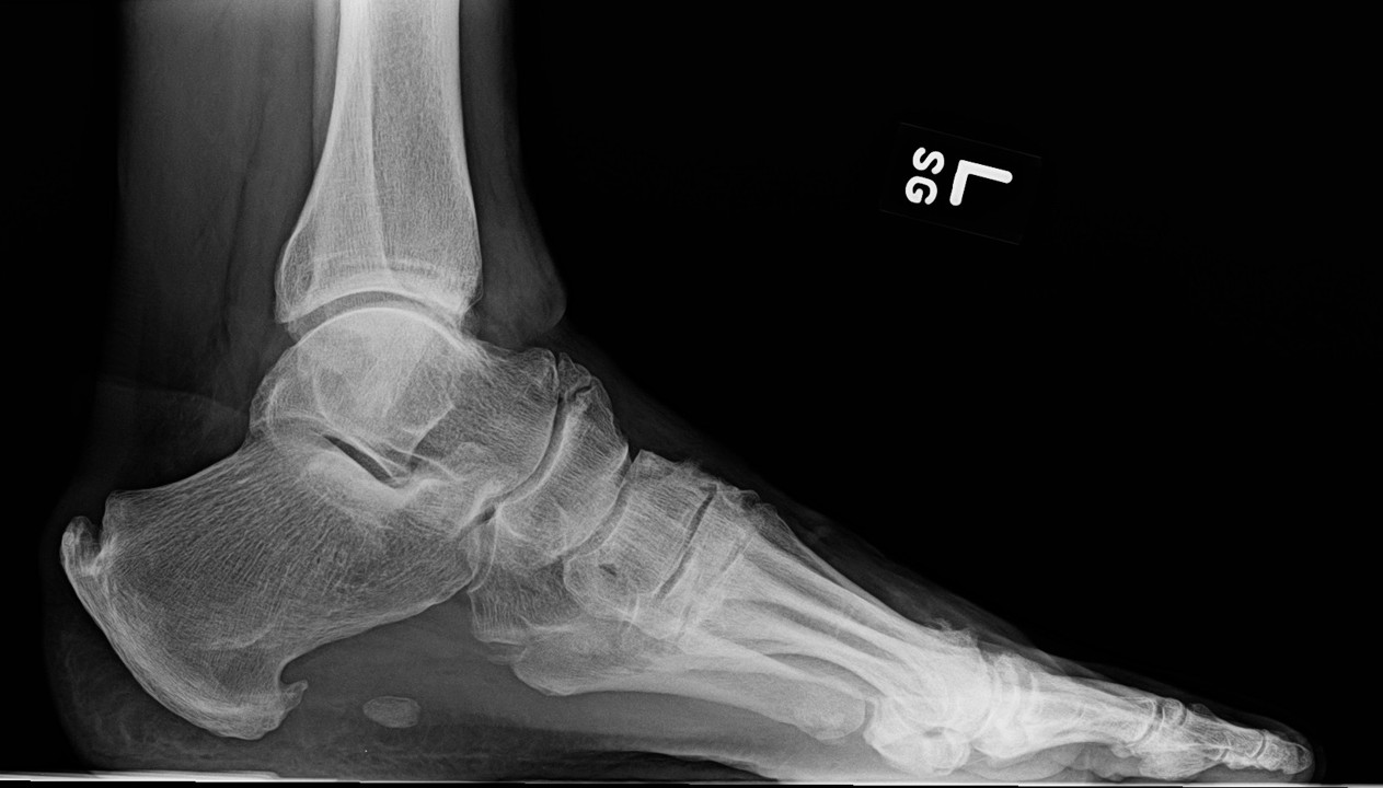
FIGURES 1: Midfoot osteophytes and joint space narrowing in left foot of 56-year-old male. (Click to enlarge.)
What Is Midfoot OA?
Midfoot osteoarthritis (OA) is a subtype of foot OA in which OA exists in one or more joints of the midfoot, most often the tarsometatarsal, talonavicular or naviculocuneiform joints (see Figure 1). OA in this region is relatively under-recognized but is a key contributor to foot pain and disability, especially in middle-to-older-aged adults.
Among adults older than 50, at least one in eight has symptomatic midfoot OA.1,2 Factors associated with symptomatic midfoot OA include older age, female sex, history of foot/ankle injury, pronated foot posture, obesity, manual occupations and pain at other weight-loaded joint sites (e.g., knees, hips).2,3 Midfoot OA shares many risk factors with other OA sites, such as the knee and hip.
Midfoot OA can occur in isolation or coexist with conditions such as rheumatoid arthritis and OA at forefoot joints. People with foot pain will often experience multi-site joint pains, which is a more likely presentation in rheumatology services.4,5 Foot pain can be overlooked during clinical exam, despite profound symptoms.6
Living with Midfoot OA?
Living with midfoot OA can be a daily struggle marked by persistent pain, stiffness or swelling. This can significantly affect mobility, making simple activities such as walking, prolonged standing, or climbing stairs difficult or exhausting.1,7,8 Discomfort often worsens during and after activity but can also occur at rest. Pain is just one aspect experienced by people living with midfoot OA. Changes in foot posture, such as flattening of the arch (Figure 2) and bony deformities, are common and can make finding suitable footwear difficult.9
Emotional well-being is often affected in people with midfoot OA, contributing to frustration, anxiety, and social withdrawal. People with midfoot OA often limit or stop social, physical and work activities, with family life and relationships also affected.10,11 These feelings are often exacerbated by a perceived lack of information about how to treat and manage midfoot OA effectively.10,11
Accessing appropriate foot care is often difficult, especially in underserved communities, due to long wait times, limited availability of foot health care and financial barriers that may delay diagnosis and treatment.12,13

FIGURES 2: Midfoot OA and flat feet (left), and midfoot flattening during heel raise (right). (Click to enlarge.)
Disparities in research amplify clinical bias, with under-representation across groups leaving gaps in knowledge and care.14 Certain populations often face systemic barriers, such as limited insurance coverage, transportation issues, and fewer culturally competent providers.15 Addressing these inequalities is essential for improving care and QoL for all people living with midfoot OA.
How Is It Evaluated Clinically?
Imaging
Radiographic diagnosis of midfoot OA has traditionally utilized the Kellgren-Lawrence system.16 More recently, the La Trobe Foot Atlas was developed to identify radiographic OA across four joints (medial and intermediate cuneiform-metatarsal joints, talonavicular joint and navicular-first cuneiform joint).17
This atlas incorporates cardinal features of OA—osteophytes and joint space narrowing—allowing for four levels of severity and diagnosis of radiographic changes (scores ≥2) plus symptoms in the corresponding region. Magnetic resonance imaging (MRI), although less accessible than X-ray, may also be used with a semiquantitative scoring system that includes all joints of the midfoot.18
Physical Exam
Midfoot OA commonly presents as persistent dorsal midfoot pain, worsening with weight-bearing and high-stress activities on the midfoot (e.g., rising onto the ball of foot, walking up or down stairs). Tight footwear pressing on the top of the midfoot may also be painful.
Pain may be reproduced with joint palpation or movement, but the proximity of the joints can make anatomical localization and diagnosis challenging without imaging.
Tarsometatarsal (Lisfranc) involvement may be identified by stabilizing the hind foot and plantarflexing the respective metatarsal (i.e., piano key test), although diagnostic accuracy is not established.19 Dorsal osteophytes may be palpable in advanced cases but limited in earlier midfoot OA.20 Localized midfoot joint swelling may be present and this is best detected with ultrasound examination.21 For unilateral symptoms, comparison to the other foot may be useful.
Midfoot pain after an axial load to a plantarflexed foot (e.g., a stumble) or following direct blunt trauma should raise suspicion of Lisfranc ligament injury or fracture, warranting urgent imaging.
If a patient has night pain, neurological symptoms, persistent inflammation and swelling or signs of infection, more serious conditions should be considered and urgent investigation undertaken.
How Do We Treat It?
Treatment of midfoot OA typically comprises biomechanical approaches, exercise and education, and pharmacological treatment.
Allied Health
A recent Australian survey showed podiatrists commonly provide footwear advice on fit, support and cushioning; use low dye taping; and prescribe arch contouring orthoses and/or shoe stiffening inserts.22 Many also advise foot muscle strengthening and, if appropriate, weight loss or referral for weight loss support.22
Evidence is limited to several case series and a randomized feasibility trial, showing immediate improvements in pain and four-week improvements in function with carbon fiber insoles, small 12-week improvements in pain with pre-fabricated arch contouring foot orthoses, and improvements in pain up to six months with custom arch-contouring foot orthoses.23-26
Although no clinical trials have tested the efficacy of the remaining approaches used in midfoot OA, strengthening exercise and weight loss are recommended in knee and hip OA clinical guidelines and thus are reasonable to consider for midfoot OA.27,28
Primary Care
Analysis of foot OA management shows physicians mainly prescribe medications, most commonly acetaminophen and non-steroidal anti-inflammatory drugs (NSAIDs).29 Although not investigated in midfoot OA, guidelines advise topical NSAIDs first for peripheral OA, and oral NSAIDs if topical preparations are ineffective or unsuitable.27 Acetaminophen is generally not recommended.19
Two case series suggest imaging-guided intra-articular glucocorticoid injections for midfoot OA are effective for up to three or four months, but not longer, aligning with OA guidelines which advise consideration of these injections when other pharmacological treatments are not successful or reasonable.27,30,31
Surgery, typically arthrodesis, is reserved for end-stage disease or recalcitrant pain, when conservative and pharmacological treatments fail.32
What New Knowledge Will Improve Midfoot OA Care?
Evidence-based care is essential to help people with painful midfoot OA maintain employment, independence and care-giving roles. To date, research has been limited. Large, rigorous, prospective, observational data and intervention trials are needed, together with agreed clinical criteria.
- Large, population-based observational studies could help clarify the natural history and modifiable risk factors of midfoot OA across diverse and under-represented groups. Identifying distinct subgroups (phenotypes) and drivers of disease may support more inclusive, patient-centered and personalized care by recognizing both local disease features and broader systemic influence.
- Large trials are needed to test the efficacy of footwear or orthoses to improve pain while standing or walking. Devices can create a firm lever or stable arch in the shoe, but their mechanisms and potential effectiveness are unclear. Improving muscle strength, which accounts for 30% variance of midfoot OA pain, is promising, but no trials have tested its treatment potential.
- Systemic aspects of midfoot OA overlap with knee and hip pain and are linked with obesity, diabetes and female sex.
- New research studies of people with midfoot OA as part of a presentation of multiple joint OA are recommended to investigate whether new weight loss solutions can improve joint pain, and also protect musculoskeletal power because muscle weakness is linked to midfoot OA and is associated with inactivity.
- A further critical gap is the lack of clinical criteria for midfoot OA. Comprehensive criteria must reflect the symptom experience and include all midfoot joints. Currently, standing radiographs detect medial and dorsal OA, but not lateral midfoot OA, despite lateral foot injuries, such as ankle sprains, being common. A large clinical data repository paired with imaging (e.g., radiographs, MRI, ultrasound) to capture morphology and disease activity, will inform clinical criteria and support optimized treatment algorithms.
In Summary
Midfoot OA is a common presentation in rheumatology services, but it may be overlooked and under-treated. People are increasingly seeking healthcare solutions for foot pain that will be attributable to OA. Future research is needed to understand the natural history and optimize management. Foot OA, especially at the midfoot, contributes to the growing burden of musculoskeletal pain, and rheumatology services are well placed to provide support.
Yvonne M. Golightly, PT, MS, PhD, is a professor and assistant dean for research at the University of Nebraska Medical Center College of Allied Health Professions, Omaha, Neb.
Jill Halstead, PhD, MRCPod, is a clinical lead for research, Leeds Community Healthcare NHS Trust, visiting research fellow and NIHR senior clinical research practitioner awardee at the University of Leeds, U.K.
John B. Arnold, PhD, a senior lecturer in the Allied Health & Human Performance Unit at the University of South Australia.
Lara Chapman, is a podiatrist and NIHR doctoral fellow at Leeds Institute of Rheumatic and Musculoskeletal Medicine, University of Leeds, U.K.
Catherine J. Bowen, PhD, BPod, is a professor of podiatry at the University of Southampton, U.K.
Marian T. Hannan, DSc, MPH, is a professor of medicine at Harvard Medical School and senior scientist at the Marcus Institute for Aging Research, Hebrew SeniorLife, Boston.
Hylton B. Menz, PhD, BPod, is a professor of podiatry at La Trobe University, Melbourne, Victoria, Australia.
Kade L. Paterson, PhD, BPod, BAppSci(Hons), is a Principal Research Fellow and a Dame Kate Campbell Fellow with the Centre for Health, Exercise and Sports Medicine in the Department of Physiotherapy at the University of Melbourne, Australia.
Martin J. Thomas, PhD, MCSP, is a senior research fellow in clinical epidemiology at Keele University and honorary specialist rheumatology and musculoskeletal physiotherapist at the Midlands Partnership University NHS Foundation Trust, U.K.
References
- Roddy E, Thomas MJ, Marshall M, et al. The population prevalence of symptomatic radiographic foot osteoarthritis in community-dwelling older adults: Cross-sectional findings from the clinical assessment study of the foot. Ann Rheum Dis. 2015 Jan;74(1):156–163.
- Thomas MJ, Peat G, Rathod T, et al. The epidemiology of symptomatic midfoot osteoarthritis in community-dwelling older adults: Cross-sectional findings from the Clinical Assessment Study of the Foot. Arthritis Res Ther. 2015 Jul 13;17(1):178.
- Arnold JB, Marshall M, Thomas MJ, et al. Midfoot osteoarthritis: potential phenotypes and their associations with demographic, symptomatic and clinical characteristics. Osteoarthritis Cartilage. 2019 Apr;27(4):659–666.
- Keenan AM, Tennant A, Fear J, et al. Impact of multiple joint problems on daily living tasks in people in the community over age fifty-five. Arthritis Rheum. 2006 Oct 15;55(5):757–764.
- Abraham L, Halsby K, Stein N, et al. An observational retrospective matched cohort study of healthcare resource utilisation and costs in UK patients with moderate to severe osteoarthritis pain. Rheumatol Ther. 2022 Jun;9(3):851–874.
- Korda J, Bálint GP. When to consult the podiatrist. Best Pract Res Clin Rheumatol. 2004 Aug;18(4):587–611.
- Menz HB, Dufour AB, Casey VA, et al. Foot pain and mobility limitations in older adults: The Framingham Foot Study. J Gerontol A Biol Sci Med Sci. 2013 Oct;68(10):1281–1285.
- Menz HB, Lord SR. Foot pain impairs balance and functional ability in community-dwelling older people. J Am Podiatr Med Assoc. 2001 May;91(5):222–229.
- Lithgow MJ, Buldt AK, Munteanu SE, et al. Structural foot characteristics in people with midfoot osteoarthritis: Cross-sectional findings from the clinical assessment study of the foot. Arthritis Care Res (Hoboken). 2024 Feb;76(2):225–230.
- Thomas MJ, Moore A, Roddy E, Peat G. “Somebody to say ‘come on we can sort this’”: A qualitative study of primary care consultation among older adults with symptomatic foot osteoarthritis. Arthritis Care Res (Hoboken). 2013 Dec;65(12):2051–2055.
- Chapman LS, Flurey CA, Richards P, et al. What outcomes are important to people with foot and ankle disorders in rheumatic and musculoskeletal diseases? An OMERACT qualitative interview study across four continents. Semin Arthritis Rheum. 2025 Jun;72:152671.
- McCulloch L, Borthwick A, Redmond A, et al. UK podiatrists’ experiences of podiatry services for people living with arthritis: A qualitative investigation. J Foot Ankle Res. 2018 Jun;11:27.
- Dando C, Bacon D, Borthwick A, Bowen C. Stakeholder views of podiatry services in the UK for people living with arthritis: A qualitative study. J Foot Ankle Res. 2020 Sep 24;13(1):58.
- Faison WE, Harrell PG, Semel D. Disparities across diverse populations in the health and treatment of patients with osteoarthritis. Healthcare (Basel). 2021 Oct 22;9(11):1421.
- Karmarkar TD, Maurer A, Parks ML, et al. A fresh perspective on a familiar problem: Examining disparities in knee osteoarthritis using a Markov Model. Med Care. 2017 Dec;55(12):993–1000.
- Kellgren JH, Lawrence JS. Radiological assessment of osteo-arthrosis. Ann Rheum Dis. 1957 Dec;16(4):494–502.
- Menz HB, Munteanu SE, Landorf KB, et al. Radiographic classification of osteoarthritis in commonly affected joints of the foot. Osteoarthritis Cartilage. 2007 Nov;15(11):1333–1338.
- Halstead J, Martín-Hervás C, Hensor EMA, et al. Development and reliability of a preliminary foot osteoarthritis magnetic resonance imaging score. J Rheumatol. 2017 Aug;44(8):1257–1264.
- Keiserman LS, Cassandra J, Amis JA. The piano key test: A clinical sign for the identification of subtle tarsometatarsal pathology. Foot Ankle Int. 2003 May;24(5):437–438.
- Thomas MJ, Roddy E, Rathod T, et al. Clinical diagnosis of symptomatic midfoot osteoarthritis: Cross-sectional findings from the Clinical Assessment Study of the Foot. Osteoarthritis Cartilage. 2015 Dec; 23(12):2094–2101.
- Camerer M, Ehrenstein B, Hoffstetter P, et al. High-resolution ultrasound of the midfoot: Sonography is more sensitive than conventional radiography in detection of osteophytes and erosions in inflammatory and non-inflammatory joint disease. Clin Rheumatol. 2017 Sep;36(9):2145–2149.
- Lim PQX, Menz HB, Landorf KB, et al. Assessment and management of midfoot osteoarthritis by podiatrists in Australia: A cross-sectional survey of current practice. Rheumatol Int. 2025 May 12;45(6):141.
- Yi T, Kim JH, Oh-Park M, Hwang JH. Effect of full-length carbon fiber insoles on lower limb kinetics in patients with midfoot osteoarthritis: A pilot study. Am J Phys Med Rehabil. 2018 Mar;97(3):192–199.
- Rao S, Baumhauer JF, Becica L, Nawoczenski DA. Shoe inserts alter plantar loading and function in patients with midfoot arthritis. J Orthop Sports Phys Ther. 2009 Jul;39(7):522–531.
- Halstead J, Chapman GJ, Gray JC, et al. Foot orthoses in the treatment of symptomatic midfoot osteoarthritis using clinical and biomechanical outcomes: A randomised feasibility study. Clin Rheumatol. 2016 Apr;35(4):987–996.
- Ibuki A, Cornoiu A, Clarke A, et al. The effect of orthotic treatment on midfoot osteoarthritis assessed using specifically designed patient evaluation questionnaires. Prosthet Orthot Int. 2010 Dec;34(4):461–471.
- Osteoarthritis in over 16s: Diagnosis and management. London: National Institute for Health and Care Excellence. 2022 Oct 19. https://tinyurl.com/6z238zpm.
- Moseng T, Vliet Vlieland TPM, Battista S, et al. EULAR recommendations for the non-pharmacological core management of hip and knee osteoarthritis: 2023 update. Ann Rheum Dis. 2024 May 15;83(6):730–740.
- Paterson KL, Harrison C, Britt H, et al. Management of foot/ankle osteoarthritis by Australian general practitioners: An analysis of national patient-encounter records. Osteoarthritis Cartilage. 2018 Jul;26(7):888–894.
- Drakonaki EE, Kho JSB, Sharp RJ, Ostlere SJ. Efficacy of ultrasound-guided steroid injections for pain management of midfoot joint degenerative disease. Skeletal Radiol. 2011 Aug;40(8):1001–1006.
- Protheroe D, Gadgil A. Guided intra-articular corticosteroid injections in the midfoot. Foot Ankle Int. 2018 Aug;39(8):1001–1004.
- Patel A, Rao S, Nawoczenski D, et al. Midfoot arthritis. J Am Acad Orthop Surg. 2010 Jul;18(7):417–425.
- Arnold JB, Halstead J, Grainger AJ, et al. Foot and leg muscle weakness in people with midfoot osteoarthritis. Arthritis Care Res (Hoboken). 2021 Jun;73(6):772–780.
- Peiris WL, Cicuttini FM, Hussain SM, et al. Is adiposity associated with back and lower limb pain? A systematic review. PLoS One. 2021 Sep;16(9):e0256720.