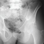
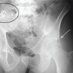
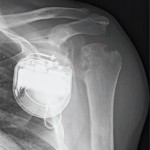
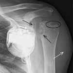
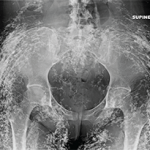
Editor’s note: In this recurring feature, we first present a series of images (this page) for your review, and then a brief discussion of the findings and diagnosis (p. 63). Before you turn to the discussion, examine these images carefully and draw your own conclusions. History For the discussion, click here. A 55-year-old man on…