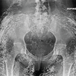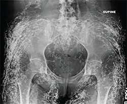
Figure 1:
Anteroposterior radiograph of the pelvis.
Findings/Diagnosis
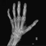
The radiographs demonstrate diffuse sheetlike and tumefactive calcifications throughout the subcutaneous tissues, muscle and fascia of the pelvis and right hand.
The underlying bones and joint spaces appear normal.
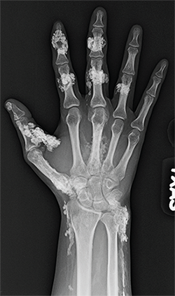
Figure 2: Anteroposterior radiograph of the right hand.
The differential diagnosis for soft tissue calcification is extensive and includes metabolic disturbances (particularly of calcium and phosphate), trauma (e.g., injection sites, tissue necrosis, resolving hematoma), collagen vascular diseases, crystal deposition disease (e.g., gout, calcium pyrophosphate and hydroxyapatite deposition disease), parasitic infections and venous insufficiency.