Video: Every Case Tells a Story| Webinar: ACR/CHEST ILD Guidelines in Practice

An official publication of the ACR and the ARP serving rheumatologists and rheumatology professionals

  • Conditions
    • Axial Spondyloarthritis
    • Gout and Crystalline Arthritis
    • Myositis
    • Osteoarthritis and Bone Disorders
    • Pain Syndromes
    • Pediatric Conditions
    • Psoriatic Arthritis
    • Rheumatoid Arthritis
    • Sjögren’s Disease
    • Systemic Lupus Erythematosus
    • Systemic Sclerosis
    • Vasculitis
    • Other Rheumatic Conditions
  • FocusRheum
    • ANCA-Associated Vasculitis
    • Axial Spondyloarthritis
    • Gout
    • Psoriatic Arthritis
    • Rheumatoid Arthritis
    • Systemic Lupus Erythematosus
  • Guidance
    • Clinical Criteria/Guidelines
    • Ethics
    • Legal Updates
    • Legislation & Advocacy
    • Meeting Reports
      • ACR Convergence
      • Other ACR meetings
      • EULAR/Other
    • Research Rheum
  • Drug Updates
    • Analgesics
    • Biologics/DMARDs
  • Practice Support
    • Billing/Coding
    • EMRs
    • Facility
    • Insurance
    • QA/QI
    • Technology
    • Workforce
  • Opinion
    • Patient Perspective
    • Profiles
    • Rheuminations
      • Video
    • Speak Out Rheum
  • Career
    • ACR ExamRheum
    • Awards
    • Career Development
  • ACR
    • ACR Home
    • ACR Convergence
    • ACR Guidelines
    • Journals
      • ACR Open Rheumatology
      • Arthritis & Rheumatology
      • Arthritis Care & Research
    • From the College
    • Events/CME
    • President’s Perspective
  • Search

Articles tagged with "diagnostic imaging"

Diagnostic Imaging in Patient with Elbow Pain: Findings

Veronika Sharp, MD, Midori Jane Nishio, MD, & Lily Kao, MD  |  May 14, 2015

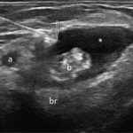
View the question. Transverse (see Figure 1) and longitudinal (see Figure 2) ultrasound images show an anechoic collection (asterisks), which surrounds the distal biceps tendon (b). The collection is compressible (not shown here). The biceps tendon is heterogeneous in echo texture, with fiber dropouts and an irregular border (in the transverse view). The elbow joint…

The Diagnostic View: Our Findings

Jennifer L. Demertzis, MD  |  April 1, 2015

Radiograph, MR images reveal symmetric erosive sacroiliitis in a patient who was diagnosed with ankylosing spondylitis

The Diagnostic View: Our Findings

Jennifer L. Demertzis, MD  |  March 1, 2015

Radiograph, MR images reveal pigmented villonodular synovitis in 54-year-old woman with right elbow pain

The Diagnostic View: Our Findings

Jennifer L. Demertzis, MD  |  February 1, 2015

Radiograph, MR images of left shoulder reveal rheumatoid arthritis in 49-year-old woman

Is Ultrasound Right For YOUR Office?

Kurt Ullman  |  July 12, 2011

Growth of ultrasound for rheumatic diseases leads many to wonder how it might fit into their practice

  • « Previous Page
  • 1
  • 2
  • 3
  • About Us
  • Meet the Editors
  • Issue Archives
  • Contribute
  • Advertise
  • Contact Us
  • Copyright © 2025 by John Wiley & Sons, Inc. All rights reserved, including rights for text and data mining and training of artificial technologies or similar technologies. ISSN 1931-3268 (print). ISSN 1931-3209 (online).
  • DEI Statement
  • Privacy Policy
  • Terms of Use
  • Cookie Preferences