Video: Every Case Tells a Story| Webinar: ACR/CHEST ILD Guidelines in Practice

An official publication of the ACR and the ARP serving rheumatologists and rheumatology professionals

  • Conditions
    • Axial Spondyloarthritis
    • Gout and Crystalline Arthritis
    • Myositis
    • Osteoarthritis and Bone Disorders
    • Pain Syndromes
    • Pediatric Conditions
    • Psoriatic Arthritis
    • Rheumatoid Arthritis
    • Sjögren’s Disease
    • Systemic Lupus Erythematosus
    • Systemic Sclerosis
    • Vasculitis
    • Other Rheumatic Conditions
  • FocusRheum
    • ANCA-Associated Vasculitis
    • Axial Spondyloarthritis
    • Gout
    • Psoriatic Arthritis
    • Rheumatoid Arthritis
    • Systemic Lupus Erythematosus
  • Guidance
    • Clinical Criteria/Guidelines
    • Ethics
    • Legal Updates
    • Legislation & Advocacy
    • Meeting Reports
      • ACR Convergence
      • Other ACR meetings
      • EULAR/Other
    • Research Rheum
  • Drug Updates
    • Analgesics
    • Biologics/DMARDs
  • Practice Support
    • Billing/Coding
    • EMRs
    • Facility
    • Insurance
    • QA/QI
    • Technology
    • Workforce
  • Opinion
    • Patient Perspective
    • Profiles
    • Rheuminations
      • Video
    • Speak Out Rheum
  • Career
    • ACR ExamRheum
    • Awards
    • Career Development
  • ACR
    • ACR Home
    • ACR Convergence
    • ACR Guidelines
    • Journals
      • ACR Open Rheumatology
      • Arthritis & Rheumatology
      • Arthritis Care & Research
    • From the College
    • Events/CME
    • President’s Perspective
  • Search

2015 ACR/ARHP Annual Meeting: Macrophage Polarization and Its Role in Inflammatory Disease

Susan Bernstein  |  Issue: April 2016  |  April 14, 2016

Fibroblast-like synoviocytes promote C9 macrophage polarization that may drive pannus formation and tissue damage in RA-affected joints, said Dr. Ivashkiv. More study of the C9 growth factor pathways may lead to better understanding of how this destruction happens in RA.

Macrophage Sub-Types

There’s little information about cell markers to identify subtypes of synovial macrophages, said Ursula Fearon, PhD, professor of molecular rheumatology at Trinity College in Dublin, Ireland.

ad goes here:advert-1
ADVERTISEMENT
SCROLL TO CONTINUE

In an RA synovium, you see more macrophages than normal. CD68 macrophages are increased in clinically inflamed knee joints, for example. “No matter the therapy, DAS 28 score changes significantly correlate to subsets of macrophages in the joints,” Dr. Fearon said.

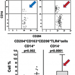
Macrophages from the synovium of active RA joints have a pro-inflammatory profile, and there is an increase in CD50 and CD36.6 In RA synovial fluid, M1 phenotypes are seen in higher amounts. Anti-citrullinated protein antibodies (ACPA) also affect how macrophage subtypes are distributed in joints, which could lead to RA, she said.7 Dr. Fearon and colleagues studied the macrophage subtypes in synovial biopsies of very inflamed joints, and also saw an increase in M1 macrophages.

ad goes here:advert-2
ADVERTISEMENT
SCROLL TO CONTINUE

“It’s difficult to define the markers of macrophage subtypes,” said Dr. Fearon. As far as targeting macrophages to develop therapies, “many patients don’t respond to these targets.”

Researchers are focusing on reprogramming to try to block the metabolic pathway in macrophages, an old idea that has come back around, she said.

GLUT-1 mediated glucose metabolism drives a pro-inflammatory macrophage phenotype. Pyruvate kinase M2, a key metabolic regulator, tetramerization inhibits Hif-1α IL-1ß, and promotes an M2 phenotype.8 “So that’s a possible therapeutic target,” she said. Metformin has also been shown to directly suppress macrophage phenotypes. So synovial macrophages clearly play a role in RA disease activity, and one day may be targeted in the joint environment as a therapy, she said.

Turning Off Macrophages

“The function of a macrophage is to protect us from infection or injury,” said Andy Clark, PhD, professor of inflammation biology at the University of Birmingham in the United Kingdom. “When it detects one of these potentially harmful events, it responds by producing pro-inflammatory cytokines like TNF.”

Normally, this response is tightly regulated, he said. The macrophage returns to its resting state and stops producing TNF. However, “in the rheumatoid synovium, it looks as though macrophages have forgotten how to switch off. We need to understand how these on and off switches work, and why they sometimes don’t operate correctly.”
Page: 1 2 3 4 5 | Single Page
Share: 

Filed under:ConditionsMeeting ReportsOther Rheumatic ConditionsResearch Rheum Tagged with:2015 ACR/ARHP Annual MeetingAmerican College of Rheumatology (ACR)DiseaseinflammationmacrophagepolarizationResearchRheumatoid arthritis

Related Articles

    Doest Rheumatoid Arthritis Take a Toll?

    November 15, 2013

    Exploring the Toll background

    New Studies Identify Possible Markers of Early Systemic Sclerosis

    August 16, 2019

    Although the true cause of systemic sclerosis (SSc), or scleroderma, remains unknown, researchers have made progress in detecting the autoimmune disease’s early presence. Beyond the physiological signs of Raynaud’s phenomenon, a capillaroscopy can detect alterations in microcirculation and lab tests can confirm the presence of telltale autoantibodies, such as anti-topoisomerase 1, anti-centromere and anti-RNA polymerase…

    Macrophage Activation Syndrome

    December 1, 2010

    A review of diagnosis, treatment, and prognosis

    ACR Winter Rheumatology Symposium: New Kinase Inhibitors Show Promise

    May 1, 2013

    Toxicity and appropriate targeting are challenges for small molecule researchers

  • About Us
  • Meet the Editors
  • Issue Archives
  • Contribute
  • Advertise
  • Contact Us
  • Copyright © 2025 by John Wiley & Sons, Inc. All rights reserved, including rights for text and data mining and training of artificial technologies or similar technologies. ISSN 1931-3268 (print). ISSN 1931-3209 (online).
  • DEI Statement
  • Privacy Policy
  • Terms of Use
  • Cookie Preferences