Early diagnosis can prevent prolonged morbidity and extensive investigations. Despite effective clinical and imaging diagnostic methods, patients can experience delays in diagnosis of months or even years, as seen with the three cases detailed above.
The three most common indicators of glomus tumors are intense paroxysmal pain, point tenderness and cold sensitivity.2,3,7
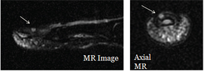
Figure 8: POCUS is used to guide an MRI study of a nail bed.
Three common clinical methods of diagnosis can be used to effectively distinguish glomus tumors from other similarly sized or located masses. Hildreth’s sign refers to the temporary relief of pain at the site of the lesion by inducing ischemia with a pressure cuff.8 Love’s pin test, in which a pinhead is used to apply pressure to the site of the tumor, should produce intense pain.9 A cold sensitivity test, which is conducted by applying cold water, an ice cube or any other material that causes a drop in temperature at the site of the lesion, should produce intense pain.10
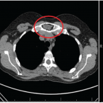
Imaging is often used to verify the diagnosis prior to surgical intervention. Standard radiographs are typically normal, but erosion of the bone can occur in some cases (as seen in Case 2).11 MRI can be most effectively used for confirming the diagnosis and the tumor’s location.10,12 MR imaging can produce false-negative results in 10% of cases. Resolution is a limiting factor, allowing small lesions to be undetected.12
Ultrasound can detect tumors as small as 2 mm in diameter, gives better visualization of tumors when compared with standard radiology techniques, and is less expensive and time consuming than MRI. Color or power Doppler ultrasound (used on Patients 2 and 3) provides enhanced vascular detail when compared with grey-scale methods.3,5,13,14
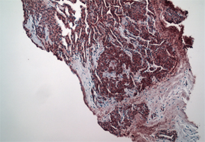
Figure 9: Smooth muscle actin immunostain shows smooth muscle surrounding vascular channels and neural elements. Courtesy of David Chercover, MD, Lions Gate Hospital, North Vancouver, B.C.
The standard course of treatment for a glomus tumor is surgical excision. Multiple tumors are reported. Recurrences are uncommon and are generally due to incomplete excision.12
Digital glomus tumors are small, rare causes of hand pain. Their small size and low prevalence make an early diagnosis difficult. As in our cases, no predisposing or risk factors are known, although the condition is three times more common in women. When faced with symptoms of paroxysmal pain, cold sensitivity and acute tenderness, physicians have clinical strategies that can be employed to identify glomus tumors with greater certainty.