Video: Every Case Tells a Story| Webinar: ACR/CHEST ILD Guidelines in Practice

An official publication of the ACR and the ARP serving rheumatologists and rheumatology professionals

  • Conditions
    • Axial Spondyloarthritis
    • Gout and Crystalline Arthritis
    • Myositis
    • Osteoarthritis and Bone Disorders
    • Pain Syndromes
    • Pediatric Conditions
    • Psoriatic Arthritis
    • Rheumatoid Arthritis
    • Sjögren’s Disease
    • Systemic Lupus Erythematosus
    • Systemic Sclerosis
    • Vasculitis
    • Other Rheumatic Conditions
  • FocusRheum
    • ANCA-Associated Vasculitis
    • Axial Spondyloarthritis
    • Gout
    • Psoriatic Arthritis
    • Rheumatoid Arthritis
    • Systemic Lupus Erythematosus
  • Guidance
    • Clinical Criteria/Guidelines
    • Ethics
    • Legal Updates
    • Legislation & Advocacy
    • Meeting Reports
      • ACR Convergence
      • Other ACR meetings
      • EULAR/Other
    • Research Rheum
  • Drug Updates
    • Analgesics
    • Biologics/DMARDs
  • Practice Support
    • Billing/Coding
    • EMRs
    • Facility
    • Insurance
    • QA/QI
    • Technology
    • Workforce
  • Opinion
    • Patient Perspective
    • Profiles
    • Rheuminations
      • Video
    • Speak Out Rheum
  • Career
    • ACR ExamRheum
    • Awards
    • Career Development
  • ACR
    • ACR Home
    • ACR Convergence
    • ACR Guidelines
    • Journals
      • ACR Open Rheumatology
      • Arthritis & Rheumatology
      • Arthritis Care & Research
    • From the College
    • Events/CME
    • President’s Perspective
  • Search

A Practical Guide to Autoantibody Testing in Rheumatic Diseases

Donald Thomas, MD, FACP, FACR, RhMSUS, & Jason Liebowitz, MD  |  Issue: October 2024  |  October 7, 2024

Several theories exist for why common autoantibodies in healthy people are nonpathogenic. Some are produced by immature B cells that are not programmed for affinity maturation by antigen stimulation or somatic mutation. Another theory is that the autoantigen is not in a location accessible to antigen-processing cells and autoreactive B cells. One method of this immune system sequestration is that many autoantigens are intracellular. Healthy apoptosis prevents immune system exposure.

ANA IFA

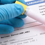
The ACR recommends ordering ANA by IFA methodology using human epidermoid tumor cell-line #2 (HEp-2) cells. Part of the reason is that ANA by IFA identifies many more possible nuclear antigen targets than the cheaper and easier to perform solid-phase immunoassays, including the multiplex immunoassay and the enzyme-linked immunosorbent assay (ELISA).8

ad goes here:advert-1
ADVERTISEMENT
SCROLL TO CONTINUE

However, several studies have demonstrated wide variations in ANA IFA results, even between instruments used by the same manufacturer.9 ANA may be positive in one laboratory, yet negative in another. Due to this, there is a push for improved ANA IFA standards among commercial laboratories.

ANA IFA Titer

The IFA ANA test applies the person’s serum to HEp-2 cells on a slide. ANAs from the patient’s blood then bind to antigens in the HEp-2 cells. The technician adds fluorescent-tagged immunoglobulins that bind to the antibodies.

ad goes here:advert-2
ADVERTISEMENT
SCROLL TO CONTINUE

Under the microscope, fluorescent-tagged ANAs appear as a yellowish-green glow. If ANAs are present, dilution tests are performed. When the technician adds an equal part of the diluting solution to the serum, the result is a 1:1 mix (or titer). When the lab technician adds a second equal amount of diluting solution, the result is a 1:2 titer and so on. This is repeated until the serum no longer fluoresces. The last titer that fluoresced is noted. Higher ANA concentrations result in higher titers.

ANA results are reported with the pattern noted by the technician as well as the titer. The titer result is the most helpful clinically. Most labs consider a titer of 1:40 or 1:80 and higher to be a positive ANA.

The greater the titer, the greater the likelihood for a systemic autoimmune rheumatic disease (SARD). An ANA of 1:80 has a positive predictive value of only 2% for a SARD, but it is around 50% with an ANA of 1:640 or higher.9

The likelihood, or post-test probability, that someone with a positive ANA has a SARD depends on the pre-test probability and the likelihood ratio of the ANA result. In clinical practice, we rarely calculate actual numbers. However, it is important to at least note subjective approximations.

Page: 1 2 3 4 5 6 | Single Page
Share: 

Filed under:Conditions Tagged with:ANA testautoantibodiesautoantibody testingEditor's Picklaboratory tests

Related Articles

    Laboratory Testing for Diagnosis, Management of Patients with Rheumatic Disease

    December 1, 2014

    A review of data on antinuclear antibodies and tests for rheumatoid arthritis

    Know Your Labs

    February 1, 2009

    A review of state-of-the-art testing for SLE and connective tissue disease.

    Can Lupus Be Prevented? Research Reveals Clues to Who’s Most Likely to Transition to Classified Disease

    February 25, 2020

    How does a patient transition from health to active SLE? This question is the crux of the research conducted by Judith A. James, MD, PhD, and colleagues…

    New Study Reveals Limitations in ANA Test Kits for Lupus

    September 20, 2018

    The variability in serological testing for antinuclear antibodies (ANA) is under investigation after unexpected findings were reported from clinical trials of new agents to treat systemic lupus erythematosus (SLE). In “Assay Variation in the Detection of Antinuclear Antibodies in the Sera of Patients with Established SLE,” David S. Pisetsky, MD, PhD, and his colleagues demonstrate…

  • About Us
  • Meet the Editors
  • Issue Archives
  • Contribute
  • Advertise
  • Contact Us
  • Copyright © 2025 by John Wiley & Sons, Inc. All rights reserved, including rights for text and data mining and training of artificial technologies or similar technologies. ISSN 1931-3268 (print). ISSN 1931-3209 (online).
  • DEI Statement
  • Privacy Policy
  • Terms of Use
  • Cookie Preferences